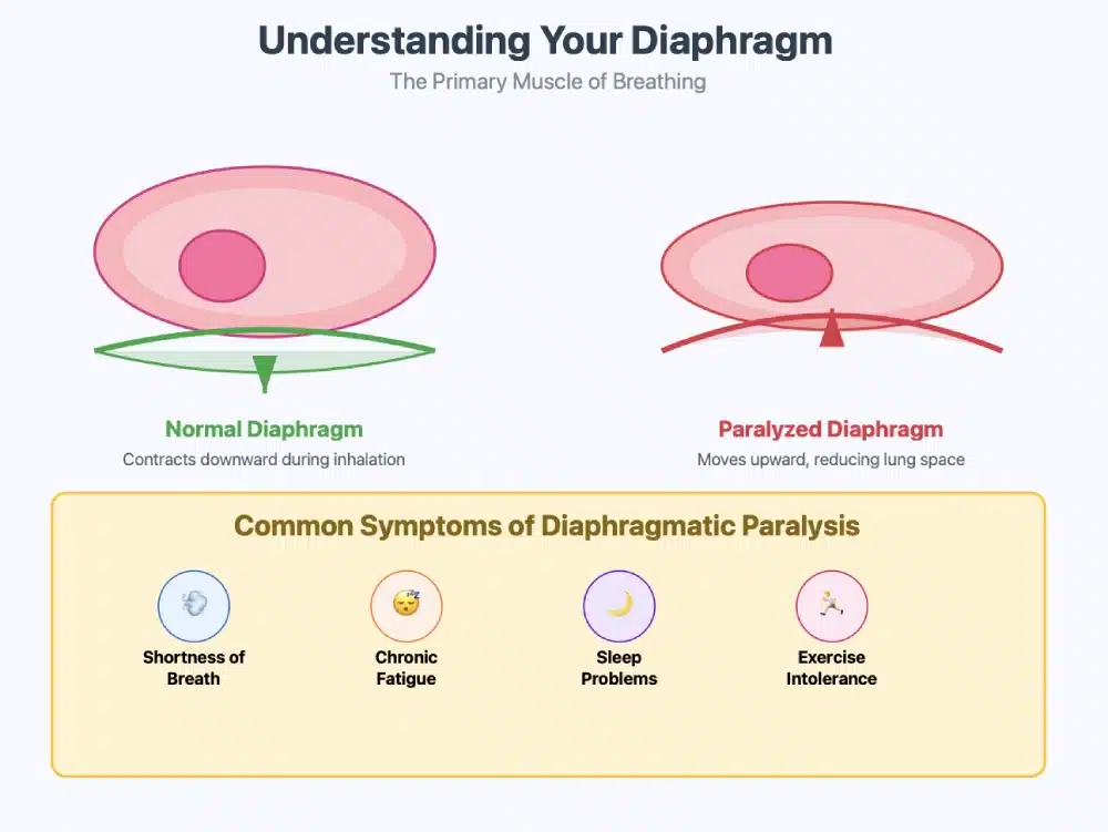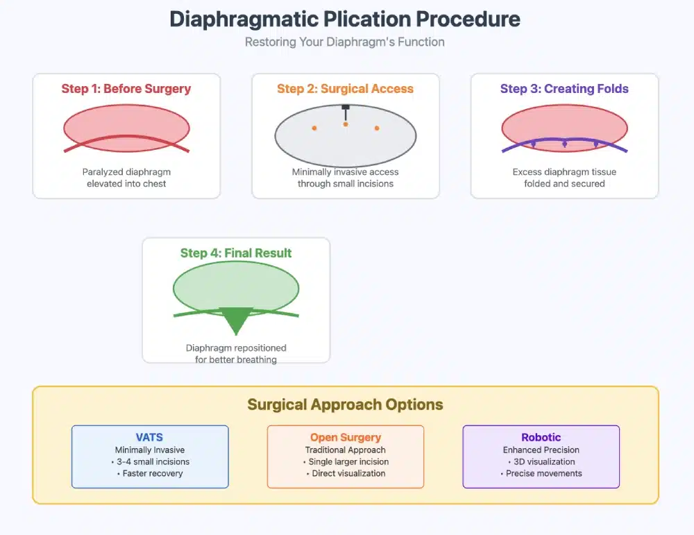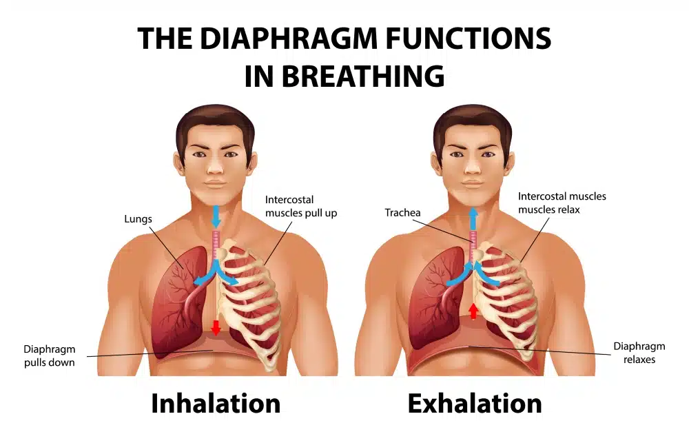
The anatomy of the diaphragm is essential for understanding the implications of its dysfunction. It is a dome-shaped muscle that separates the thoracic cavity from the abdominal cavity, playing a pivotal role in the mechanics of breathing. When the diaphragm becomes weakened or paralyzed, it can result in poor lung expansion and inadequate ventilation, which are often seen in patients suffering from chronic respiratory diseases or post-surgical complications. It effectively addresses these issues by surgically folding and suturing the diaphragm in its functional position.

What is it?
It refers to a surgical procedure aimed at correcting functional abnormalities of the diaphragm, a crucial muscle involved in respiration. In cases where the diaphragm is paralyzed, often due to conditions such as diaphragmatic paralysis or a hernia, the muscle cannot contract effectively, leading to compromised respiratory function. The purpose is to restore normal diaphragm position and function, thereby improving lung capacity and the overall respiratory process.
How to Treat Diaphragm Dysfunction
When the diaphragm is unable to contract properly, it can lead to significant respiratory issues. This dysfunction can result from a variety of causes including neurological conditions, trauma, obesity, and certain chronic diseases such as COPD or asthma. The impact of diaphragm dysfunction extends beyond impaired respiration; it can also affect overall health by causing complications such as reduced oxygenation of tissues, fatigue, and decreased exercise capacity. Furthermore, in severe cases, diaphragm dysfunction can lead to respiratory failure, requiring medical intervention to support breathing.
There are several types of diaphragm dysfunction, including paradoxical diaphragm movement, which occurs when the diaphragm moves in an incorrect direction during inspiration. This can be particularly noticeable in individuals who have suffered from nerve injury or other conditions affecting the diaphragm. The prevalence of diaphragm dysfunction varies among different populations, often being more common in patients with underlying pulmonary conditions or those who have experienced trauma. Additionally, the condition is frequently observed in the elderly, who may face a decline in muscle strength and function, leading to an increased risk of diaphragm-related issues. Understanding diaphragm dysfunction and its implications is essential for identifying suitable treatment options and improving respiratory health.
Overview of the Surgical Procedure
The procedure is generally performed under general anesthesia and can be executed through various techniques, including open surgery or minimally invasive approaches such as thoracoscopic surgery. Patients typically experience improved diaphragm stability, enhanced lung function, and a reduction in respiratory distress. Moreover, this intervention has been found particularly beneficial for individuals with significant diaphragm dysfunction, as it can lead to an overall improvement in quality of life, enabling better physical activity and daily functioning.
Why Consider it?
Unilateral Diaphragmatic Paralysis due to Phrenic Nerve Injury
The phrenic nerve, which plays a vital role in diaphragmatic function, can be compromised by various causes, leading to unilateral diaphragm paralysis. One of the primary causes is traumatic injury, which can occur from blunt or penetrating trauma to the neck or thoracic area. Such trauma can sever or damage the phrenic nerve directly, resulting in disruptions to its signaling capabilities. Furthermore, sports injuries or accidents may also lead to significant nerve damage, necessitating careful diagnosis and treatment for affected individuals.
Surgical complications represent another leading cause of phrenic nerve injury. Procedures involving the thoracic or cervical regions, such as cardiac surgeries or cervical spine surgeries, can inadvertently affect the phrenic nerve due to its anatomical proximity. Surgeons must maintain a high level of awareness regarding the nerve’s location to mitigate risks during these operations.
In addition to trauma and surgery, tumors in the neck or thoracic region can compress the phrenic nerve, leading to dysfunction. Both benign and malignant growths can exert pressure on the nerve, impairing its ability to control the diaphragm effectively. Conditions such as lung cancer or metastatic disease can be particularly concerning as they may lead to notable impairments of respiratory function.
Inflammatory or degenerative diseases, such as amyotrophic lateral sclerosis (ALS) or multiple sclerosis, are also recognized for their potential to affect the phrenic nerve. These conditions can lead to a progressive weakening of the nerve and, consequently, diaphragmatic paralysis. Identifying these underlying health conditions is critical for intervention and management. Very rarely viral infections might be the cause as well.
Diaphragm Eventration and Elevated Hemidiaphragm
Diaphragm Eventration refers to an abnormal elevation of the diaphragm due to the thinning or weakness of the muscle fibers, resulting in a part of the diaphragm being pushed upward into the chest cavity. This condition is often congenital but can also occur secondary to trauma, surgery, or certain neuromuscular diseases. The weakened area can lead to respiratory symptoms such as shortness of breath, especially with exertion, and can be associated with gastrointestinal issues if the abdominal organs herniate into the chest.
Elevated Hemidiaphragm occurs when one side of the diaphragm (hemidiaphragm) is raised above its normal position. This can be due to a variety of causes, including phrenic nerve paralysis, lung diseases (like pneumonia, atelectasis, or pleural effusion), or after abdominal surgery. An elevated hemidiaphragm can cause restrictive lung patterns, leading to reduced lung volumes and impaired respiratory function. Symptoms are often nonspecific but can include dyspnea (difficulty breathing) and chest discomfort.
Addressing Respiratory Failure and Diaphragmatic Hernia
In patients with both respiratory failure and a diaphragmatic hernia, the hernia may worsen the respiratory condition by compressing the lungs and impairing normal ventilation. Addressing the hernia often requires surgical repair, either via open surgery or minimally invasive techniques like laparoscopy, to reposition the herniated organs and close the defect. Managing respiratory failure in these cases typically involves stabilization with mechanical ventilation and supportive care until the hernia is surgically corrected.
Diagnosing Diaphragm Issues
Several diagnostic tools are employed to accurately assess diaphragmatic function and understand the extent of paralysis. One prevalent method involves utilizing imaging studies; chest x-ray or chest radiography or chest radiograph can reveal an elevated hemidiaphragm, indicative of paralysis. In more complex cases, a CT scan or magnetic resonance imaging (MRI) may be required to visualize the phrenic nerve and depict any lesions or abnormalities that might be contributing to the paralyzed diaphragm.
Additionally, healthcare practitioners may perform a physical examination, focusing on assessing diaphragmatic movement through techniques such as palpation or ultrasonography. This is done sitting up and in supine position. These methods can provide real-time feedback on diaphragmatic excursion during respiration. The combination of clinical evaluation techniques, along with advanced imaging and function testing, allows for an accurate diagnosis of unilateral diaphragmatic paralysis, ensuring appropriate management and treatment for affected individuals.
Pulmonary Function Tests
Pulmonary function tests (PFTs) play a critical role in evaluation, measuring the lung’s capacity to move air in and out. In patients with diaphragmatic paralysis, these tests typically reveal a restrictive pattern, accompanied by reduced inspiratory volume in the affected hemithorax. Furthermore, electromyography (EMG) may be utilized to assess the electrical activity of the diaphragm and detect any neuromuscular involvement. EMG findings can confirm whether the paralysis results from a primary nerve lesion or secondary muscular degeneration.
Measuring Level of Dyspnea and Dyspnea Scores
Dyspnea, or difficulty in breathing, is a common and distressing symptom experienced by individuals with respiratory or cardiovascular conditions. Accurately measuring and assessing the level of dyspnea is critical for diagnosing underlying causes, guiding treatment decisions, and monitoring the progression of diseases. Various tools and scoring systems have been developed to quantify dyspnea, helping clinicians assess its severity and impact on a patient’s quality of life.
Dyspnea Scales and Scores
- The Borg Dyspnea Scale (Borg Scale)
One of the most commonly used tools for measuring dyspnea is the Borg Rating of Perceived Exertion (RPE) Scale, which includes a section specifically for dyspnea. The scale ranges from 0 to 10, where:- 0 represents “nothing at all” (no dyspnea),
- 10 represents “maximum possible” (severe, distressing dyspnea).
This scale allows patients to rate their perceived difficulty in breathing based on their subjective experience. It is widely used in clinical settings, especially during exercise tests or physical activities, to assess exertional dyspnea.
- The Modified Medical Research Council (mMRC) Dyspnea Scale
The mMRC Dyspnea Scale is a simple, self-reported tool used to assess the severity of dyspnea in individuals with chronic respiratory conditions like Chronic Obstructive Pulmonary Disease (COPD). It consists of five grades, ranging from:- Grade 0 (no dyspnea except on strenuous exercise) to
- Grade 4 (dyspnea prevents leaving the house or occurs at rest).
This scale is easy to administer and is helpful for gauging the functional impact of dyspnea on daily activities.
- The Visual Analog Scale (VAS)
The Visual Analog Scale is a straightforward tool in which patients mark their level of dyspnea on a continuous 100mm line, where one end represents “no dyspnea” and the other represents “the worst possible dyspnea.” This tool is particularly useful for capturing fine gradations in severity and allows for a quantitative measure that can be tracked over time. - The Dyspnea-12 (D-12) Scale
The Dyspnea-12 Scale is a more detailed assessment tool, consisting of 12 items that evaluate both the physical and emotional aspects of dyspnea. It is designed to be self-reported and provides a more comprehensive view of how dyspnea affects the patient’s daily life, including questions related to breathlessness during exertion, rest, and sleep. - The Shortness of Breath Questionnaire (SOBQ)
The SOBQ is often used in patients with chronic respiratory diseases such as COPD, interstitial lung disease, or heart failure. This scale assesses both the frequency and severity of breathlessness during activities of daily living, providing a score that helps clinicians evaluate a patient’s dyspnea over time.
Surgical Techniques for Treatment of Diaphragmatic Paralysis – Plication Procedures in Adult Patients
Open Thoracotomy Approach or Open Surgery (Traditional Approach):
The traditional surgical treatment for repairing a diaphragmatic hernia involves a large abdominal incision or thoracoabdominal approach. The surgeon repositions the herniated organs back into the abdominal cavity, repairs the defect in the diaphragm with sutures, and may reinforce the repair with mesh if necessary to prevent recurrence.
Video-Assisted Thoracoscopic Surgery (VATS) or thoracoscopic approach
Laparoscopic Plication (Minimally Invasive) or Minimally Invasive Diaphragmatic Plication
Laparoscopic techniques are becoming increasingly popular for diaphragmatic hernia repair due to their minimal invasiveness and quicker recovery time. Small incisions are made, and the surgeon uses a camera (laparoscope) to guide the repair. Mesh may also be used for reinforcement, but the laparoscopic method results in less postoperative pain and shorter hospital stays.
The Role of General Anesthesia in Diaphragmatic Surgery
Diaphragmatic surgery is a complex and delicate procedure that requires precise management of both the surgical site and the patient’s respiratory function. General anesthesia plays a critical role in ensuring the success of diaphragmatic repairs, such as those for diaphragmatic hernia, diaphragmatic rupture, diaphragmatic eventration, and other related conditions. This article explores the essential role of general anesthesia in diaphragmatic surgery, its benefits, and key considerations for anesthesiologists during these procedures.
What Is General Anesthesia?
General anesthesia is a medically induced state of unconsciousness, with the primary goal of providing analgesia (pain relief), muscle relaxation, and amnesia (memory loss) during surgical procedures. For diaphragmatic surgery, general anesthesia ensures that the patient is completely unconscious, immobile, and unaware of the surgery, while also protecting the airway and optimizing respiratory function.
Key Roles of General Anesthesia in Diaphragmatic Surgery
- Ensuring Patient Comfort and Safety
Diaphragmatic surgeries, such as repairing diaphragmatic hernia or rupture, can be particularly painful and involve complex maneuvers. General anesthesia ensures that patients do not experience pain, discomfort, or distress during the operation. Additionally, it maintains complete control over the patient’s airway, which is critical when working near vital respiratory structures. - Managing Airway and Ventilation
One of the main challenges in diaphragmatic surgery is the manipulation of the diaphragm, which directly impacts breathing mechanics. During surgery, the diaphragm may be temporarily displaced or impaired, and general anesthesia allows anesthesiologists to secure the airway with an endotracheal tube or laryngeal mask airway. This enables controlled mechanical ventilation, ensuring that oxygen levels remain stable, and the patient’s breathing is carefully regulated throughout the procedure.Mechanical ventilation is essential because patients undergoing diaphragmatic surgery may already have compromised respiratory function due to conditions such as COPD or diaphragmatic eventration. General anesthesia, combined with ventilatory support, helps maintain adequate oxygenation and carbon dioxide elimination. - Facilitating Muscle Relaxation
Diaphragmatic surgery often requires complete muscle relaxation to allow for precise surgical access, especially in cases involving complex hernia repairs or repairs of the diaphragm following trauma. Muscle relaxants administered during general anesthesia provide the necessary relaxation, which makes it easier for the surgeon to work around the diaphragm and surrounding structures, such as the lungs, heart, and abdominal organs. - Controlling Hemodynamics and Stress Response
Surgery on the diaphragm may trigger a stress response in the body, leading to increased heart rate, blood pressure, and potentially dangerous fluctuations in cardiac output. General anesthesia helps control these physiological responses by administering appropriate anesthetic agents and vasodilators, ensuring that the patient’s cardiovascular system remains stable throughout the surgery. - Protecting the Diaphragm and Respiratory Function
For surgeries involving the repair of a diaphragmatic hernia or rupture, general anesthesia is used to minimize the risk of complications related to respiratory function. The anesthesiologist continuously monitors end-tidal carbon dioxide (EtCO2) levels and oxygen saturation to ensure that adequate gas exchange occurs, especially when the diaphragm is temporarily rendered nonfunctional during the procedure.Additionally, lung-protective ventilation strategies are employed to prevent barotrauma (injury caused by excessive pressure) and atelectasis (lung collapse) when managing the patient’s ventilation during surgery. This is crucial, especially for patients with underlying pulmonary conditions. - Postoperative Pain Management
Effective pain control is essential following diaphragmatic surgery, as the diaphragm is a highly sensitive muscle involved in the respiratory cycle. General anesthesia allows for regional anesthesia techniques, such as epidural blocks or paravertebral blocks, which can provide extended pain relief post-surgery. By combining general anesthesia with regional techniques, anesthesiologists can reduce opioid requirements, thereby minimizing the risk of side effects such as nausea, vomiting, and respiratory depression.
Benefits
Improved Respiratory Function and Lung Capacity
One of the most significant benefits of surgery is its ability to restore proper diaphragm function. A weakened or elevated diaphragm severely restricts lung expansion and ventilation, leading to dyspnea (shortness of breath) and poor oxygen exchange. By shortening and repositioning the diaphragm, plication helps the muscle regain its normal ability to contract, significantly improving lung function and oxygenation.
Long-Term Improvement in Pulmonary Function
For patients with diaphragmatic eventration or paralysis, breathing difficulties can be persistent and debilitating. Plication reduces the degree of diaphragm dysfunction, leading to immediate improvement in respiratory symptoms. Many patients report relief from chronic shortness of breath, progressive dyspnea and an enhanced ability to engage in daily activities without feeling fatigued or breathless.
Enhanced Quality of Life and Daily Activities
Patients who experience significant dyspnea or respiratory distress often face limitations in their daily lives, including difficulty exercising, performing physical tasks, and even resting comfortably. By improving diaphragm function, it helps restore normal breathing mechanics, enabling patients to breathe more easily and engage in physical activity. This leads to a marked improvement in quality of life and overall well-being.
Reducing Shortness of Breath
For patients with diaphragmatic eventration or paralysis, breathing difficulties can be persistent and debilitating. Plication reduces the degree of diaphragm dysfunction, leading to immediate improvement in respiratory symptoms. Many patients report relief from chronic shortness of breath and an enhanced ability to engage in daily activities without feeling fatigued or breathless.
Risks and Complications

Pleural Effusion and Infections
Infections are a potential risk following any surgery. Infection can occur at the surgical site, particularly if the incisions are not properly cleaned or if bacteria enter the body during the procedure. Infections may lead to increased pain, swelling, and the need for further treatment, including antibiotics or, in severe cases, a second surgery to drain abscesses.
- Risk factors: Poor hygiene, smoking, or underlying conditions like diabetes can increase the risk of infection.
Bleeding and Hemorrhage
Bleeding is another potential complication, especially during the manipulation of the diaphragm and surrounding tissues. While bleeding is usually minimal in minimally invasive surgeries, it can be more significant in cases of open surgery or when the diaphragm is severely damaged.
- Management: Surgeons take great care to minimize bleeding, but in some cases, blood transfusions or additional procedures may be needed to control significant blood loss.
Injury to Surrounding Organs
The diaphragm is located near several vital organs, including the lungs, heart, liver, and stomach. There is a risk of accidental injury to these structures, particularly when working near the phrenic nerve or the abdominal cavity. Although rare, such injuries can lead to complications like lung collapse, pneumothorax, or damage to the liver or stomach.
- Preventive measures: Surgeons use advanced imaging techniques and minimally invasive methods to reduce the risk of organ damage.
Pneumothorax (Collapsed Lung)
In laparoscopic or robotic surgery, pneumothorax (air in the chest cavity) is a potential complication. This can occur if air or gas used during the surgery leaks into the pleural space, leading to a collapsed lung. Pneumothorax can result in chest pain, difficulty breathing, and the need for immediate medical intervention, such as chest tube insertion to remove the trapped air.
- Risk factors: Pneumothorax is more common in patients with pre-existing lung conditions or in cases where the diaphragm has been weakened by disease or trauma.
Nerve Injury
The phrenic nerve, which controls diaphragm movement, is closely associated with the diaphragm muscle. There is a risk of inadvertent injury to the phrenic nerve, which can worsen diaphragmatic dysfunction or cause paralysis of the diaphragm. Nerve injury can lead to ongoing breathing difficulties, shortness of breath, and the need for additional treatments or surgeries.
- Management: Surgeons take extreme care when performing the procedure near the phrenic nerve to minimize the risk of injury.
Postoperative Pain and Recovery Time
While most patients experience some level of pain following surgery the degree of discomfort varies depending on the approach used (open vs. minimally invasive). Patients may experience muscle soreness or discomfort from the surgical site, which can interfere with normal activities during the recovery phase.
- Postoperative pain management: Doctors often recommend pain relievers such as NSAIDs or opioids, along with regional anesthesia techniques (like epidural blocks) to manage pain during recovery.
Scarring and Adhesions
In any abdominal or thoracic surgery there is a risk of scarring and the formation of adhesions (bands of fibrous tissue that bind organs together). Adhesions can lead to chronic pain, bowel obstruction, or other complications if they interfere with normal organ function.
- Minimizing scarring: Minimally invasive techniques are less likely to result in significant scarring compared to traditional open surgery.
Recurrence of Diaphragmatic Dysfunction
Although this surgery generally offers long-term relief, in some cases, the condition may recur, especially if the underlying cause (e.g., neurological issues or trauma) is not fully addressed. This can result in the reappearance of breathing difficulties, necessitating a second surgery or alternative treatments.
- Long-term monitoring: Patients are typically monitored postoperatively to ensure that the diaphragm remains functional and that no recurrence occurs.
Anesthesia Risks
As with any surgery, general anesthesia carries inherent risks, particularly for patients with pre-existing health conditions like cardiovascular disease or respiratory disorders. Complications from anesthesia may include allergic reactions, breathing difficulties, or heart arrhythmias.
Patients might require Mechanical Ventilation
- Preoperative assessment: A thorough evaluation by the anesthesiologist ensures that patients are properly prepared for the procedure and that risks are minimized.
Chest Tube Requirements
All patients get a small chest drain after surgery to ensure drainage of air and fluid and avoid collection in the chest.
Pulmonary Embolism
This is a blood clot in the blood vessels that go to the lung, it can be serious and make patients very short of breath.
Who is a Candidate for This Type of Surgery?
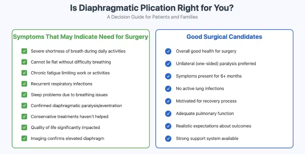
Not every patient with diaphragmatic dysfunction is a suitable candidate for surgery. The ideal candidates are those who experience significant respiratory distress due to diaphragm dysfunction, particularly when other treatments (such as respiratory therapy or ventilatory support) have not provided sufficient relief. Here are some of the primary factors that determine whether a patient is a good candidate for the procedure:
1. Patients with Diaphragmatic Eventration and treatment of diaphragmatic eventration
Diaphragmatic eventration occurs when part of the diaphragm becomes abnormally elevated and loses its ability to contract properly. This condition can result in breathing difficulties, poor oxygenation, and chronic shortness of breath. Surgery is particularly effective for patients with unilateral diaphragmatic eventration (affecting one side of the diaphragm), as it helps restore the diaphragm’s normal position and improves respiratory function.
- Key symptoms: Chronic shortness of breath, fatigue, difficulty performing physical activities, and impaired lung function.
- Ideal candidates: Patients with moderate to severe diaphragmatic eventration who do not respond to conservative treatments like ventilator support or respiratory therapy.
2. Patients with Diaphragmatic Paralysis
Diaphragmatic paralysis, often caused by nerve damage (e.g., from trauma, surgery, or neurological conditions like stroke or polio), results in one or both sides of the diaphragm failing to function properly. Surgery is often recommended for patients with unilateral diaphragmatic paralysis (paralysis of one side of the diaphragm) who experience severe breathing difficulties and reduced quality of life. By restoring the diaphragm’s function, plication can improve the patient’s ability to breathe without relying on mechanical ventilation.
- Key symptoms: Severe shortness of breath, difficulty breathing during exertion, and reliance on ventilators.
- Ideal candidates: Patients with unilateral diaphragmatic paralysis who have not responded to conservative treatments and those who require improved lung expansion for normal respiration.
3. Patients with Diaphragmatic Weakness Due to Neurological conditions or diaphragmatic palsy.
Certain neurological conditions can cause diaphragmatic weakness, including neuromuscular diseases like muscular dystrophy and amyotrophic lateral sclerosis (ALS). For patients whose diaphragmatic weakness significantly impairs breathing, surgery can help restore diaphragm function and improve overall respiratory mechanics.
- Key symptoms: Progressive difficulty breathing, shortness of breath, and compromised lung capacity.
- Ideal candidates: Patients with neurological conditions that lead to diaphragmatic weakness, particularly those with unilateral diaphragm involvement.
4. Patients with Post-Traumatic Diaphragmatic Dysfunction
Traumatic injury to the diaphragm (e.g., from trauma, surgery, or penetrating injuries) can result in diaphragmatic dysfunction, including elevated diaphragm or diaphragmatic hernia. If the damage results in impaired diaphragm function, plication can be used to correct the diaphragm’s position and improve respiratory efficiency.
- Key symptoms: Difficulty breathing, chest pain, and limited lung expansion.
- Ideal candidates: Patients with diaphragmatic dysfunction resulting from trauma who experience significant respiratory distress and have not improved with conservative treatments.
5. Patients Who Are Not Candidates for More Complex Surgery
In some cases, patients with severe diaphragmatic dysfunction may be considered candidates when they are not suitable for more complex surgeries such as diaphragmatic transplant or lung surgery. Plication can offer a simpler, less invasive alternative to restore diaphragm function and reduce symptoms.
- Key symptoms: Persistent shortness of breath, difficulty breathing during normal activities, and limited ability to exercise or perform physical tasks.
- Ideal candidates: Patients with severe diaphragmatic dysfunction who are not suitable candidates for more invasive procedures and only one affected side.
Additional Considerations for Surgery
Surgery is highly effective for many patients, but certain factors may influence whether a patient is a suitable candidate for the procedure:
1. Overall Health Status
- General health: Patients should be in good general health and capable of tolerating surgery. This includes having stable cardiovascular function, manageable blood pressure, and normal lung function.
- Comorbid conditions: Conditions such as severe pulmonary disease, heart disease, or severe obesity may affect the decision to proceed with plication, as they can increase the risks associated with surgery.
2. Type and Extent of Diaphragmatic Dysfunction
- Unilateral vs. bilateral involvement: surgery is typically more effective for patients with unilateral (one-sided) diaphragm dysfunction, as the other side may still maintain some function.
- Severity: Patients with severe diaphragmatic dysfunction, especially those who have developed compensatory mechanisms (such as relying on the remaining healthy lung), may benefit most from plication.
3. Age and Life Expectancy
While surgery can be performed on patients of various ages, younger and healthier patients are generally better candidates for surgery. Age and life expectancy can influence the decision to proceed with plication, especially if the underlying cause of diaphragmatic dysfunction is progressive (e.g., neuromuscular disease).
Patient Selection for Symptomatic Patients
Patient selection is critical when considering surgery, as not all individuals will benefit from the procedure. Factors such as the underlying reason for diaphragmatic dysfunction, overall health status, and comorbid conditions must be weighed. Additionally, individual variations in anatomy and physiology can significantly influence surgical outcomes, necessitating a thorough preoperative assessment to gauge potential efficacy and risks.
Recovery expectations must also be managed carefully; patients may need to engage in a structured rehabilitation program following surgery to regain optimal lung function and strength. Therefore, understanding the potential risks of surgery is essential for both patients and healthcare providers to make informed decisions and ensure a balanced view of the procedure’s pros and cons.
Recovery and Aftercare
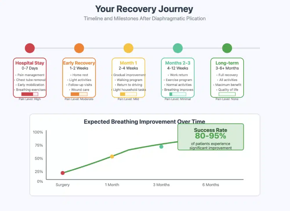
The recovery process largely depends on the surgical approach used (minimally invasive or open surgery), the patient’s overall health, and the extent of the diaphragmatic dysfunction. Here’s what patients can generally expect during their recovery:
1. Hospital Stay
- Minimally invasive surgery (laparoscopy or robotic-assisted) typically requires a shorter hospital stay, often 1 to 2 days, while open surgery may require a stay of 3 to 5 days.
- During the hospital stay, patients will be monitored for vital signs, including breathing, heart rate, and oxygen levels.
- Pain management will be provided, and any drains or tubes used during the surgery will be monitored and removed as necessary.
2. Immediate Postoperative Period
After the surgery, patients may experience:
- Soreness or mild discomfort in the chest or abdominal area, which is common after diaphragm surgery.
- Fatigue and weakness as the body heals.
- A temporary reduction in lung function as the diaphragm adjusts to its new position and function.
- Respiratory monitoring may continue, particularly if the patient had severe breathing difficulties before surgery.
3. Gradual Return to Normal Activities
- Week 1-2: Most patients can expect to feel fatigued and may have difficulty moving or performing simple activities. During this time, it is crucial to rest and avoid physical exertion.
- Week 3-4: Patients may begin to feel less short of breath and can gradually resume light activities. It’s important to follow the surgeon’s instructions for physical activity to avoid straining the diaphragm.
- Week 4-6: By this time, many patients begin to feel more energized and can return to non-strenuous work or daily activities.
Pain Management and Medications
Managing pain is an important part of the recovery process. Your doctor will prescribe pain medications, including NSAIDs (non-steroidal anti-inflammatory drugs) or opioids if needed. Many patients also benefit from:
- Local anesthetics or nerve blocks are administered during or after surgery to manage postoperative pain.
- Ice packs or cold compresses to reduce swelling and discomfort in the first few days after surgery.
- Breathing exercises: These exercises help improve lung function and ease discomfort associated with shallow breathing.
Physical Activity and Mobility
Following surgery, it is essential to give the body time to heal before resuming normal physical activity. However, it is also important to avoid long periods of inactivity, as this can lead to complications such as blood clots and muscle weakness. Here’s how to approach activity levels during the recovery period:
- Week 1-2: Rest is critical in the initial phase, but light walking and gentle movements (as tolerated) are encouraged to promote circulation and prevent blood clots.
- Week 3-4: Gradually increase activity levels with light stretching and walking. Avoid lifting heavy objects or engaging in strenuous exercises.
- Month 1-2: After the initial recovery phase, most patients can slowly reintroduce more intense activities, but high-impact sports or heavy lifting should still be avoided.
Diet and Nutrition During Recovery
Proper nutrition plays a vital role in supporting the body’s healing process after surgery. Here are some key considerations:
- Hydration: Drink plenty of fluids to stay hydrated and support lung function.
- Protein-rich foods: Include lean meats, fish, eggs, and plant-based proteins to promote tissue repair and muscle strength.
- Small, frequent meals: Especially during the first few days post-surgery, eating smaller, more frequent meals can help manage discomfort from nausea or abdominal bloating that sometimes occurs after surgery.
Follow-Up Appointments
- Postoperative visits are essential to ensure that the diaphragm is healing properly and functioning as expected.
- Your healthcare professionals will monitor lung function, assess breathing improvement, and adjust your treatment plan if necessary.
- Chest X-rays or CT scans may be performed to assess the position of the diaphragm and ensure there are no complications.
Long-Term Recovery and Outcomes
A review of the literature shows that most patients experience significant improvement in respiratory function and overall quality of life after diaphragmatic plication. Long-term recovery includes:
- Improved lung capacity and better oxygenation, leading to less shortness of breath and increased activity tolerance.
- Increased independence from mechanical ventilation or oxygen support, if previously required.
- Restoration of normal diaphragm function, allowing for more effective breathing during physical activity.
- Long term improvement of pulmonary function
Long term results of diaphragmatic plication and Outcomes – physiologic results of diaphragm plication.
The success rate of surgical plication is generally high, with most patients experiencing significant improvement in respiratory function and quality of life following surgery. Success is measured by the restoration of diaphragm function, reduction in breathing difficulties, and the ability to engage in normal activities without the need for mechanical ventilation or supplemental oxygen.
1. Improvement in Respiratory Function
The main goal is to restore proper diaphragm movement and improve lung function. Case series have shown that 90% to 95% of patients experience a marked improvement in lung volumes, oxygenation, and overall respiratory efficiency. Patients typically report less shortness of breath and a significant reduction in the need for ventilator support.
2. Reduced Reliance on Mechanical Ventilation
For patients who have been using a ventilator or positive pressure ventilation due to diaphragmatic dysfunction, diaphragmatic plication can often reduce or eliminate the need for mechanical ventilation. Approximately 80% to 85% of patients who underwent diaphragmatic plication for diaphragmatic paralysis or eventration no longer required mechanical ventilation after recovery.
3. Long-Term Success
Long-term success rates are also favorable, with studies showing that 85% to 90% of patients maintain improved respiratory function for years following surgery. This indicates that diaphragmatic plication is not only effective in the short term but also provides sustained benefits for most patients.
Factors That Affect Success Rates
1. Type of Diaphragmatic Dysfunction
- Diaphragmatic eventration: Patients with unilateral diaphragmatic eventration (one-sided diaphragm dysfunction) tend to have better outcomes compared to those with bilateral diaphragmatic dysfunction.
- Diaphragmatic paralysis: The success in patients with diaphragmatic paralysis(caused by nerve damage) depends on the extent of nerve injury and whether other neurological conditions are present. Unilateral paralysis typically leads to better outcomes than bilateral paralysis.
2. Timing of Surgery
The timing of surgery can significantly impact success rates. Early intervention after a diagnosis of diaphragmatic dysfunction is associated with better results. Delaying surgery can lead to muscle atrophy or permanent damage to the diaphragm, making surgery less effective.
3. Surgical Approach
The approach used for the surgery—laparoscopic, robotic-assisted, or open surgery—can also influence success rates. Minimally invasive approaches typically result in quicker recovery times, fewer complications, and better long-term outcomes, but may not be suitable for all patients, particularly those with complex diaphragmatic dysfunction.
4. Patient’s Overall Health
Patients in good overall health tend to have better outcomes after surgery. Those with pre-existing pulmonary diseases (e.g., COPD, asthma) or cardiovascular conditions may experience slower recovery or reduced improvement in lung function. Additionally, the age of the patient can play a role, with younger and healthier patients often having faster recovery times and more significant improvements.
5. Presence of Other Conditions
Patients who have neurological diseases (e.g., muscular dystrophy, ALS) or other underlying health conditions that affect the diaphragm’s function may see less dramatic improvement after surgery. These patients may also need ongoing management to address their underlying conditions, even after plication.
Why Choose Mr. Marco Scarci
Expertise in General Thoracic Surgery: I have great experience in this operation and I carry it out mostly by keyhole approach, occasionally by open surgery in complex cases.
Specialization in Video-Assisted Thoracic Surgery: I completed a fellowship in advanced minimally invasive surgery in Canada and I have been a pioneer in the introduction of single-cut surgery in the UK.
Personalized Care for London Patients: thanks to telemedicine I can offer you my expertise everywhere in the world and get you to London only for the actual surgery.
Frequently Asked Questions
What are the Cost and Insurance Options?
1. Surgery Type (Minimally Invasive vs. Open Surgery)
- The cost for keyhole surgery typically ranges from £15,000 to 40,000, including surgeon fees, anesthesia, and hospital charges. Robotic-assisted surgery can be more expensive due to the advanced technology involved, with costs ranging from £25,000 to 50,000.
- Open surgery, which may require a longer hospital stay and intensive care unit stay, can cost between £30,000 to 60,000 or more, depending on the complexity of the surgery.
2. Surgeon’s Fees
The surgeon’s fees will vary based on their level of expertise and geographic location. Surgeons in urban areas or those with specialized expertise in thoracic surgery or diaphragmatic disorders may charge more for their services. These fees can range from £5,000 to £15,000, depending on the complexity of the case and the surgeon’s experience.
3. Anesthesia Fees
Anesthesia fees are another factor in the total cost of the procedure. For general anesthesia, the cost can range from £1,000 to 3,000, depending on the duration of the surgery and the anesthesiologist’s fee.
5. Preoperative and Postoperative Care
- Preoperative evaluations (including imaging tests like X-rays, CT scans, or MRI scans) may add £500 to 3,000to the total cost of the surgery.
- Postoperative care, including follow-up visits, medication, and possible physical therapy, may cost an additional £1,000 to £5,000.
Is Surgery Right for You?
It is an effective treatment for patients with diaphragmatic dysfunction, including conditions like diaphragmatic eventration, paralysis, and weakness. The procedure is particularly beneficial for patients who experience severe respiratory distress and have not found relief from conservative treatments like ventilatory support or respiratory therapy.
If you are struggling with breathing difficulties due to diaphragmatic dysfunction, consult with a thoracic surgeon or pulmonary specialist to determine whether this type of surgery is the right solution for you. Your healthcare provider will evaluate your overall health, the extent of diaphragmatic dysfunction, and other factors to ensure that the surgery provides the best possible results.