

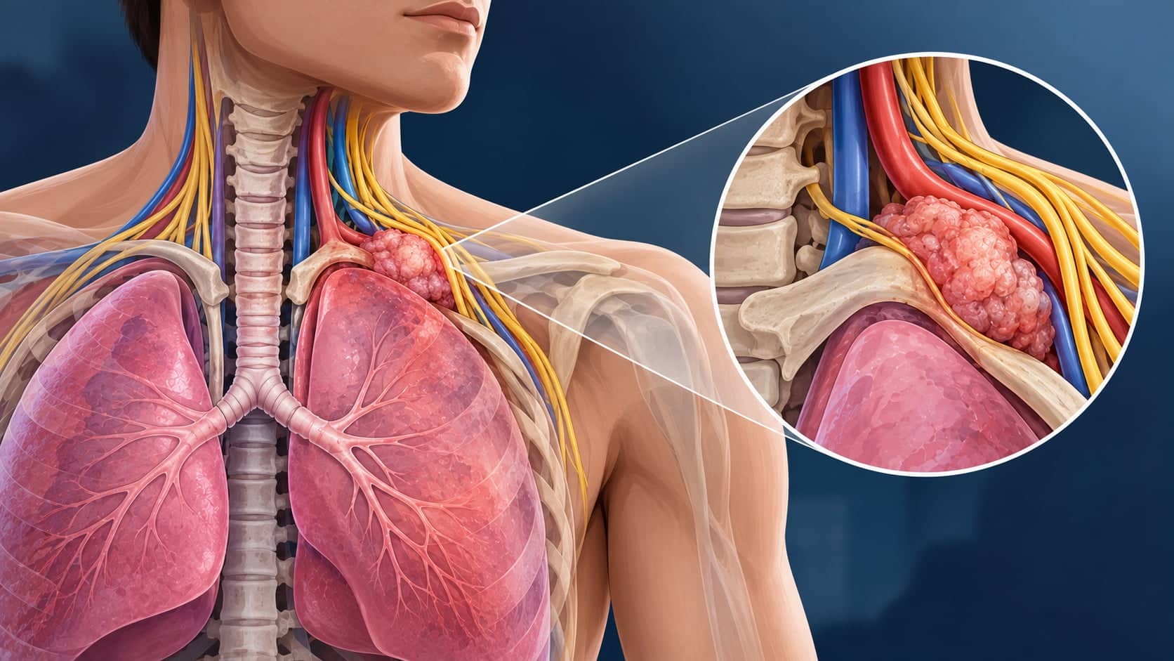
Pancoast tumours are rare lung cancers that arise at the very top of the lung, where they can involve the ribs, brachial plexus, major blood vessels, and upper spine. Because they often cause shoulder pain, arm pain, hand weakness or Horner’s syndrome rather than typical respiratory symptoms, diagnosis can be delayed.
This guide explains how Pancoast tumours are recognised and staged, what determines surgical resectability, and how modern chemoradiotherapy and surgery are combined in selected patients.
A Pancoast tumour is a rare type of lung cancer that arises at the very top, or apex, of the lung, accounting for less than 5% of lung cancer cases. Most are non-small cell lung cancer, typically adenocarcinoma or squamous cell carcinoma.
Pancoast syndrome causes severe shoulder and arm pain, hand weakness, and Horner’s syndrome due to invasion of the brachial plexus, ribs, and sympathetic chain rather than typical breathing symptoms such as cough or breathlessness.
Accurate staging with CT, MRI, and PET-CT is essential to determine whether a Pancoast tumour is surgically resectable and to plan trimodality therapy with induction chemoradiotherapy followed by surgery.
Most resectable Pancoast tumours treated today receive induction chemoradiotherapy followed by complex en bloc surgical resection performed by specialist thoracic surgeons. Video-assisted thoracoscopic surgery (VATS) or hybrid approaches may be used in selected patients.
What Are Pancoast Tumours and Pancoast Syndrome?
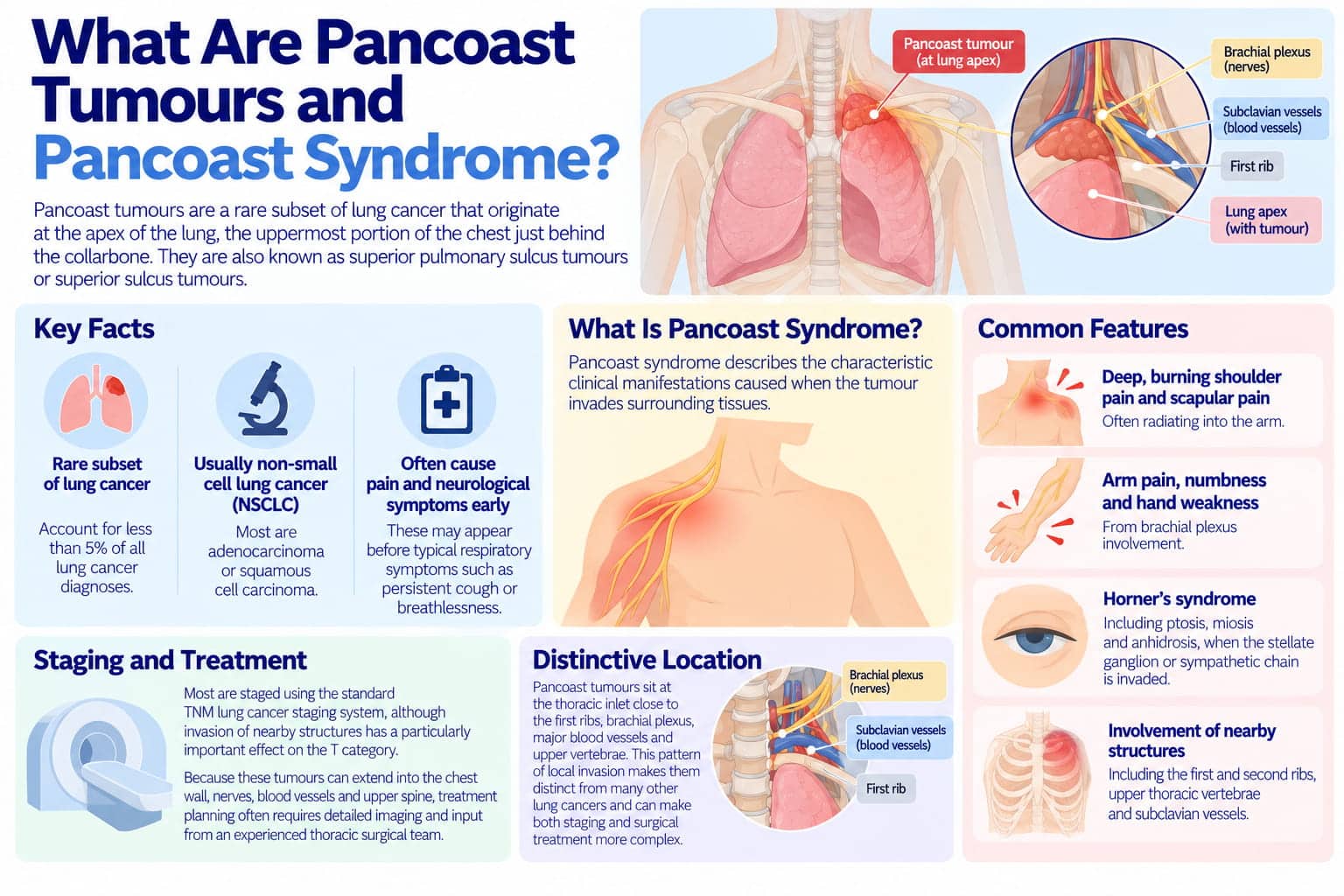
Pancoast tumours are a rare subset of lung cancer that originate at the apex of the lung, the uppermost portion of the chest just behind the collarbone. Also known as superior pulmonary sulcus tumours or superior sulcus tumours, they sit at the thoracic inlet close to the first ribs, brachial plexus, major blood vessels and upper vertebrae.
Most Pancoast tumours are non-small cell lung cancer (NSCLC), usually adenocarcinoma or squamous cell carcinoma, and they account for less than 5% of lung cancer diagnoses. Unlike more central lung tumours, they often cause pain and neurological symptoms before typical respiratory symptoms such as persistent cough or breathlessness.
Pancoast syndrome describes the characteristic clinical manifestations caused when the tumour invades surrounding tissues. These can include:
Deep, burning shoulder pain and scapular pain, often radiating into the arm;
Arm pain, numbness, and hand weakness from brachial plexus involvement;
Horner’s syndrome, including ptosis, miosis, and anhidrosis, when the stellate ganglion or sympathetic chain is invaded;
Involvement of chest wall structures including the first and second ribs, upper thoracic vertebrae, and subclavian vessels.
This pattern of local invasion is what makes Pancoast tumours distinct from many other lung cancers and can make both staging and surgical treatment more complex. Most are staged using the standard TNM lung cancer staging system, although invasion of nearby structures has a particularly important effect on the T, or tumour, category.
Because these tumours can extend into the chest wall, nerves, blood vessels and upper spine, treatment planning often requires detailed imaging and input from an experienced thoracic surgical team. Mr Marco Scarci is a London-based consultant thoracic surgeon who manages complex lung cancer, including tumours involving the lung apex and surrounding thoracic structures, and uses minimally invasive techniques where appropriate.
Want a specialist opinion on your condition?
Get a clear answer from a surgeon who will be with you throughout.
Risk Factors and Epidemiology
Pancoast tumours share many risk factors with other forms of lung cancer but have some epidemiological nuances worth noting.
The majority develop in current or former smokers, often with a significant smoking history. Risk also increases with age, particularly in older adults. Additional risk factors can include occupational exposure to asbestos, silica, and certain industrial fumes, prior chest radiotherapy, and family history of lung cancer.
Pancoast tumours account for a rare subset of NSCLC, representing less than 5% of lung cancer cases. Both men and women can be affected, although historical series included more men because of previous differences in smoking prevalence.
Because Pancoast tumour cases often present with musculoskeletal and neurological symptoms rather than cough or breathlessness, diagnosis may be delayed as symptoms resemble musculoskeletal problems. Shoulder arthritis, rotator cuff disease and cervical spine disorders can initially appear more likely than a lung tumour.
Symptoms and Early Clinical Clues
Recognising early, often non-respiratory Pancoast tumour symptoms is critical to diagnosing these cancers before they become inoperable. The most common early symptom is severe shoulder and arm pain, not cough or breathlessness.
Typical pain often begins as a deep, aching pain at the shoulder tip, scapula, or inner upper arm and may radiate down the ulnar side of the forearm into the fourth and fifth fingers. It is often worse at night and may not improve with routine musculoskeletal treatments. Neck pain or chest pain can also develop.
Neurological features can include numbness or tingling in the hand or forearm, focal weakness or clumsiness, difficulty with fine motor tasks, and wasting of the small hand muscles. These signs arise because of brachial plexus invasion by the tumour.
Horner’s Syndrome
Horner syndrome may occur with Pancoast tumours, and its presence should prompt urgent thoracic imaging. It includes a drooping eyelid, constricted pupil, and reduced sweating on the affected side of the face.
Horner syndrome is reported in approximately 20% of Pancoast tumour cases.
Common symptoms of “classic” lung cancer, such as persistent cough, coughing up blood and significant breathlessness, are often absent in the early stages. This is why Pancoast tumours may initially be misdiagnosed as shoulder arthritis, rotator cuff injury, or cervical spine disease.
Persistent or unexplained shoulder pain should prompt medical evaluation, particularly in an older patient with progressive unilateral neurological symptoms and a history of smoking.
Not sure about your treatment options?
Mr Scarci provides expert consultations typically within one week of contact.
Diagnostic Workup and Imaging
Pancoast tumour diagnosis usually requires a combination of clinical suspicion, high-quality imaging, and tissue biopsy to confirm the lung cancer type.
Limitations of Chest X-Ray
A chest X-ray can miss Pancoast tumours because of their apical location. The upper chest can be partly obscured by the clavicle, ribs and surrounding structures, meaning abnormalities may be subtle or non-specific despite significant symptoms.
Main Imaging Modalities
Modality | Role | Notes |
Contrast-enhanced CT | Visualise lung apex, chest wall invasion, rib destruction | CT has historically shown sensitivity of about 60% for defining local extent. |
MRI of thoracic inlet | Assess brachial plexus, soft tissue, spinal canal invasion | MRI has reported sensitivity around 88% for assessing local tumour extent |
PET-CT | Detect lymph node involvement, distant metastases, aid staging | Particularly important for N- and M-staging |
CT and MRI are crucial for diagnosing and assessing Pancoast tumours. Historical comparative data reported approximately 60% sensitivity for CT versus 88% for MRI when assessing the local extent of Pancoast tumours.
Each modality contributes to assessing structures critical to surgical resectability, including the vertebral bodies, spinal canal, brachial plexus roots, subclavian artery and vein, and first and second ribs. PET-CT adds important information about mediastinal lymph nodes and distant metastases.
Tissue Diagnosis
Needle biopsies are commonly used to diagnose Pancoast tumours, with CT-guided percutaneous biopsy achieving diagnostic yields above 90% in published series. Video-assisted thoracic surgery (VATS) may allow direct visualisation and biopsy when the tumour cannot be adequately sampled through a percutaneous approach.
All imaging and biopsy results should be discussed in a multidisciplinary team (MDT) involving thoracic surgeons, medical oncologists, radiation oncologists, radiologists and pathologists. The goal is to confirm the diagnosis, determine stage and decide on the most appropriate treatment plan.
"After two years of being told my symptoms were anxiety, Mr Scarci identified the problem immediately. The surgery was straightforward and I was discharged after three days. I only wish I had found him sooner."
Pathology and Lung Cancer Subtypes in Pancoast Tumours
Most Pancoast tumours are non-small cell lung cancers. Histology and molecular testing now influence not just prognosis but also possible targeted treatment options.
The main histological subtypes include adenocarcinoma, commonly reported in contemporary series; squamous cell carcinoma, historically the predominant subtype; and large cell carcinoma and other NSCLC variants, which are less frequent. Small cell carcinoma can rarely present as a Pancoast tumour and is usually treated with systemic therapy and radiotherapy rather than upfront surgery.
Once a biopsy is obtained, the pathology team may perform immunohistochemistry and molecular profiling to identify clinically relevant biomarkers such as EGFR, ALK, ROS1, KRAS, and PD-L1 expression. These findings can influence targeted therapy or immunotherapy options depending on stage and treatment setting.
The pathological assessment after induction chemoradiotherapy and surgery, including the degree of tumour response, margin status and nodal involvement, is an important predictor of long-term outcome. Patients achieving a pathological complete response (pCR) generally have more favourable outcomes.
Have a question about your diagnosis?
A consultation gives you a personalised plan — not a generic estimate.
Staging of Pancoast Tumours and Assessment of Resectability
Pancoast tumours are staged using the standard TNM lung cancer staging system, with T describing tumour size and local invasion, N describing lymph node involvement, and M describing distant metastases. Accurate staging underpins every decision about surgery.
T-Category Considerations
The T stage is influenced not only by size but also by invasion of nearby structures. Lung cancer invading the chest wall, ribs, vertebral bodies, brachial plexus, subclavian vessels or spinal structures can significantly affect the T category.
Superior sulcus tumours invading more extensive neighbouring structures may be classified as T4, while many Pancoast tumours are diagnosed at T3 or T4 because of their inherent proximity to the thoracic inlet.
N-Staging
PET-CT, endobronchial ultrasound (EBUS), EUS or mediastinoscopy may be used to evaluate hilar and mediastinal lymph nodes. Extensive mediastinal nodal disease, particularly persistent N2 or N3 disease, is an important adverse prognostic factor and can limit the role of surgery.
M-Staging
Distant metastases in sites such as the brain, liver, adrenal glands, or bone change the treatment strategy towards systemic therapy and other non-surgical approaches. Absence of distant spread, or M0 disease, is generally essential when considering a curative surgical approach.
Surgical resectability means the ability to achieve complete R0 removal of the tumour and involved chest wall structures with an acceptable functional risk. Key factors considered by thoracic surgeons and MDTs include:
Degree of brachial plexus involvement, particularly whether disease extends above the lower roots;
Spinal canal or spinal cord involvement;
Encasement of the subclavian artery or vein and whether vascular reconstruction is feasible;
The patient’s lung function and overall fitness;
Mediastinal nodal status;
Response to induction chemoradiotherapy.
Current surgical literature considers invasion of the brachial plexus above the C8 root and significant spinal canal or spinal cord involvement among important contraindications to resection, while selected vascular and vertebral involvement may be operable in highly specialised centres.
Modern pooled data show an R0 resection rate of approximately 87.6% among surgically treated patients selected after staging and multimodality therapy.
"I had rib pain for 6 years, was bounced around NHS without diagnosis. I was recommended to Mr Scarci — at consultation within minutes he knew what the problem was. Was offered surgery date the next week. 2 weeks post surgery I am almost back to full health."
Modern Treatment Strategy: Trimodality Therapy
Treatment of Pancoast tumours has evolved from historically inoperable disease to modern trimodality therapy combining chemotherapy, radiotherapy and surgery in carefully selected patients.
For resectable, non-metastatic disease, treatment may include neoadjuvant chemotherapy and radiotherapy before surgery. A typical historical regimen has involved platinum-based induction chemotherapy given concurrently with radiotherapy, followed by restaging and en bloc surgical resection when the disease remains operable.
Trimodality therapy, with induction chemoradiotherapy followed by surgery, remains a guideline-supported treatment approach for appropriate T3-T4, N0-N1 superior sulcus NSCLC.
The landmark SWOG 9416 trial demonstrated a five-year survival rate of approximately 44% overall and 54% among patients achieving complete resection.
Additional treatment after surgery depends on the pathological findings, previous systemic treatment, molecular profile and current oncology recommendations. Decisions between surgery plus chemoradiation and definitive chemoradiation alone depend on tumour stage, resectability, comorbidities and patient preference.
In Mr Marco Scarci’s multidisciplinary practice, treating Pancoast tumours involves close coordination between thoracic surgery, oncology, radiology, pathology and anaesthesia.
Want a specialist opinion on your condition?
Get a clear answer from a surgeon who will be with you throughout.
Surgical Approaches and Techniques for Pancoast Tumours

Surgery for Pancoast tumours is complex because of their proximity to critical structures and should be performed in centres with expertise in thoracic inlet anatomy. Surgery often requires techniques tailored to the exact location and extent of invasion.
Surgical Goal
A Pancoast tumour resection performed with curative intent usually involves lobectomy, most often upper lobectomy, combined with en bloc removal of involved chest wall structures.
Surgery may involve removing part of the lung and ribs, including the first and sometimes second or third ribs, parts of vertebral transverse processes, and occasionally involved segments of the subclavian vessels or brachial plexus roots.
Main Open Surgical Approaches
The high posterolateral thoracotomy, or Paulson approach, is commonly used for predominantly posterior tumours. Anterior approaches such as the Dartevelle transclavicular or transmanubrial approach provide access to the subclavian vessels and anterior ribs, while combined or “double” incisions may be needed when both anterior vascular control and posterior chest wall removal are required.
Minimally Invasive and Hybrid Techniques
Video-assisted thoracoscopic surgery (VATS) can be used for the lobectomy and lymph node dissection component in selected patients. Video-assisted thoracoscopy combined with a limited incision for chest wall resection represents a hybrid approach that may reduce the surgical trauma associated with the pulmonary part of the procedure.
Not all Pancoast tumours are suitable for purely keyhole approaches. However, when oncologically appropriate, minimally invasive components may be incorporated into a complex resection.
Mr Scarci specialises in minimally invasive lung surgery and may use VATS, robotic or hybrid techniques in selected patients, depending on the location and extent of the Pancoast tumour.
Radiotherapy, Systemic Treatment and Targeted Options
Most Pancoast tumours are treated with a combination of local therapies and systemic therapies tailored to the individual patient.
Radiotherapy Roles
Radiotherapy may be used for:
Induction chemoradiotherapy to reduce tumour burden and improve the potential for complete resection;
Post-operative radiotherapy in selected situations according to previous treatment, margins and MDT recommendations;
Palliative radiotherapy for pain relief when surgery is not possible, or disease is metastatic.
Chemotherapy
Standard induction regimens have historically used platinum-based combinations, including cisplatin and etoposide. Their purpose is to treat both the visible tumour and microscopic cancer cells that may exist beyond the primary mass.
Immunotherapy
Immunotherapy may be a treatment option for selected patients with locally advanced, recurrent or metastatic NSCLC depending on stage, biomarkers, previous treatment and current guidelines. PD-1/PD-L1-directed therapies form an important part of modern lung cancer treatment, although the optimal integration of immunotherapy specifically into Pancoast tumour trimodality pathways continues to evolve.
A recent systematic review noted that trimodality therapy remains the guideline-endorsed standard for resectable superior sulcus tumours, while evidence specifically addressing immunotherapy within these regimens remains limited.
Targeted Therapy
If the tumour harbours specific driver alterations such as EGFR, ALK or ROS1, targeted treatment may be considered depending on the stage and treatment setting. Molecular testing on biopsy tissue helps identify whether these therapies may be relevant.
The oncology team and thoracic surgeon should explain the benefits, side effects and goals of each treatment component before therapy begins. Patients undergoing resection can also prepare for lung cancer surgery recovery as part of the wider treatment plan.
Not sure about your treatment options?
Mr Scarci provides expert consultations typically within one week of contact.
Outcomes, Prognosis and Long-Term Follow-Up
Pancoast tumours were once considered uniformly lethal, but survival outcomes have improved substantially with modern multimodality treatment in experienced centres.
Key Outcome Figures
Metric | Value |
5-year survival, modern pooled data | ~49.9% |
5-year survival after complete resection in SWOG 9416 | 54% |
Pathological complete response rate | ~31.9% |
30-day surgical mortality | ~3.8% |
Pooled R0 resection rate | ~87.6% |
Complete resection with negative margins remains one of the most important favourable prognostic factors. Important prognostic factors also include the absence of significant mediastinal nodal involvement, good pathological response to induction chemoradiotherapy, absence of distant metastases, extent of brachial plexus, spinal and vascular involvement, and overall pathological stage.
Tumour-related nerve damage may affect long-term quality of life, including residual shoulder stiffness, arm weakness or sensory change. Physiotherapy, occupational therapy and structured pain management can help improve function after treatment.
Follow-up typically includes regular clinical review and interval imaging, with the exact schedule determined by tumour stage, treatment, pathology and local lung cancer surveillance protocols. Brain imaging may also be considered where symptoms or disease characteristics indicate increased risk.
Individualised survivorship care, including smoking cessation, pulmonary rehabilitation and monitoring for treatment side effects, is an important part of long-term management.
When to Seek Specialist Thoracic Surgical Advice
Early referral to a thoracic surgeon is important when a Pancoast tumour is suspected because modern multimodality treatment means some tumours previously considered inoperable can now be resected in carefully selected patients.
Situations where prompt specialist input is advisable include:
Persistent unilateral shoulder or arm pain with neurological symptoms in a current or former smoker;
Imaging showing a mass in the lung apex;
Unexplained destruction of the first rib or upper thoracic vertebra;
Confirmed or strongly suspected Pancoast tumour on CT, MRI or PET-CT;
Uncertainty about whether a superior sulcus mass is surgically resectable.
Patients who have been told that a tumour is inoperable may also seek a lung cancer second opinion so that imaging, pathology, staging and potential surgical options can be reviewed again.
Mr Scarci offers private thoracic surgery pathways in London as well as NHS practice and virtual consultations for UK and international patients who wish to discuss diagnosis, staging, or possible surgery for Pancoast tumours or other lung cancers.
Have a question about your diagnosis?
A consultation gives you a personalised plan — not a generic estimate.
Living With and After Pancoast Tumour Treatment
A Pancoast tumour diagnosis and its treatment can have a significant physical and emotional impact. Understanding what to expect during recovery helps patients plan ahead and regain confidence, particularly after complex lung and chest wall surgery.
Common post-treatment challenges include pain and stiffness in the shoulder and chest wall after rib resection, reduced lung function and breathlessness after lobectomy, and ongoing numbness or weakness in the arm if nerves were affected or partially sacrificed.
Structured rehabilitation may include physiotherapy to restore range of motion and reduce chest wall stiffness, breathing exercises and pulmonary rehabilitation, occupational therapy for daily tasks and fine motor function, and tailored exercise programmes to safely rebuild stamina. Psychological support, counselling and peer support may also be helpful, and family members or carers can play an important role in recovery.
In Mr Scarci’s practice, follow-up after surgery focuses not only on scans and tests but also on quality of life, pain control, return to work, driving, travel, and the gradual resumption of normal activities. His guidance on post-surgery recovery provides additional information about rehabilitation after lung cancer surgery.
Report any new or persistent symptoms, such as new pain, cough, weight loss, or neurological change, promptly, as they may warrant earlier review or further investigation.
FAQs About Pancoast Tumours
Are Pancoast tumours always a form of lung cancer?
The term “Pancoast tumour” usually refers to a lung cancer (almost always NSCLC) arising in the superior pulmonary sulcus at the lung apex. However, other rare tumours – such as metastases from distant cancers, lymphomas, or chest wall sarcomas – can occur in the same region and mimic Pancoast tumours on thoracic imaging. A tissue biopsy is therefore essential to confirm the lesion is truly a primary lung cancer and to guide appropriate treatment.
Can Pancoast tumours be cured?
Cure is possible in selected patients whose Pancoast tumours are localised (no distant spread), respond well to induction chemoradiotherapy, and can be completely removed surgically with clear margins. While some patients live many years disease-free, others may experience recurrence – particularly in the brain. Long-term surveillance with regular imaging and clinical review is always required.
How long does recovery from Pancoast tumour surgery usually take?
The initial hospital stay is typically five to ten days in uncomplicated cases, followed by several weeks of gradual recovery at home, a pattern similar to other thoracic operations such as pneumothorax surgery and pleurodesis. Many patients take eight to twelve weeks before returning to light work or normal daily activities. Full recovery of shoulder and arm function can take longer, especially if chest wall and nerves were involved. Early physiotherapy and adherence to rehabilitation exercises significantly influence outcomes.
Is keyhole (VATS) surgery an option for all Pancoast tumours?
While video-assisted thoracoscopic surgery can be used for parts of the operation – such as lobectomy or lymph node dissection – the need to remove involved ribs and chest wall often still requires an open or hybrid approach. Suitability depends on tumour size, position, extent of chest wall and vascular involvement, and the experience of the surgical team. The overriding priority is always complete and safe cancer removal.
Can I get a second opinion about Pancoast tumour staging and surgery?
Seeking a second opinion for a rare and complex condition is entirely reasonable and often helpful for peace of mind. Mr Marco Scarci offers in-person and virtual second-opinion consultations, during which he reviews imaging, pathology, and current treatment plans, and advises whether additional staging tests or alternative surgical strategies should be considered.
