

A collapsed lung can strike without warning, leaving you struggling for breath and unsure of what has happened. Whether triggered by a chest injury, an underlying lung condition or seemingly nothing at all, understanding the causes helps you get the right treatment and reduce the risk of it happening again.
Most cases of a collapsed lung (pneumothorax) result from air leaking into the space between the lung and chest wall. Others stem from blocked airways (atelectasis) or severe chest trauma.
Symptoms often begin suddenly with sharp chest pain and shortness of breath and can become life-threatening if not treated urgently.
Treatment ranges from observation and extra oxygen to needle aspiration, chest drain insertion and minimally invasive surgery, depending on the size and cause of the collapse.
The main risk factors include smoking and vaping, underlying lung disease such as COPD or emphysema, being tall and slim, a previous pneumothorax, chest injuries and certain genetic conditions.
Understanding Collapsed Lung: Pneumothorax and Atelectasis
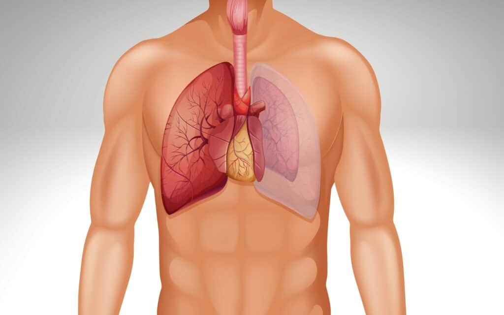
The term “collapsed lung” usually refers to a pneumothorax, where air escapes into the pleural space, the thin gap between the lung and chest wall. This extra air disrupts the negative pressure that normally keeps the lung expanded, causing it to partially or fully collapse.
A collapsed lung can also mean atelectasis, where part of the lung deflates from the inside due to a mucus plug, tumour or shallow breathing after surgery, without air entering the pleural space. Both conditions reduce oxygen levels and strain the heart and remaining lung tissue, particularly in older patients or those with pre-existing lung disease.
In extreme cases, a tension pneumothorax can develop: trapped air builds pressure inside the chest cavity, compressing the heart and major blood vessels. This is immediately life-threatening and requires emergency treatment.
The rest of this article covers the main causes of lung collapse, followed by symptoms, diagnosis and treatment options, including when referral to a thoracic specialist is advisable.
Want a specialist opinion on your condition?
Get a clear answer from a surgeon who will be with you throughout.
The Main Types of Pneumothorax and Their Causes
Pneumothorax is the most common reason for a collapsed lung. It can arise spontaneously, from injury, as a complication of a medical procedure, or as a result of underlying lung disease.
Primary spontaneous pneumothorax occurs without obvious injury, most often in otherwise healthy, tall, thin people aged roughly 20 to 40. Small air blisters called blebs or bullae on the lung surface rupture, allowing air to leak into the pleural space. Smoking, vaping and cannabis use significantly increase the risk, and recognising spontaneous pneumothorax signs and treatment is important for early care. In England, primary spontaneous pneumothorax affects approximately 15.5 per 100,000 young men per year.
Secondary spontaneous pneumothorax happens in people with known lung disease such as COPD, emphysema, cystic fibrosis, pulmonary fibrosis or lung cancer, making it vital to understand the common symptoms and warning signs of pneumothorax. Already weakened lung tissue tears more easily, meaning even a cough or minor physical strain can trigger a collapse.
Traumatic pneumothorax results from direct chest injury such as road traffic accidents, sports collisions, falls or penetrating wounds such as a knife or gunshot injury. Rib fractures can puncture the lung, causing rapid air leakage into the chest cavity.
Iatrogenic pneumothorax is an accidental complication of medical procedures such as central line insertion, lung biopsy, mechanical ventilation or deep acupuncture. Studies show roughly 1.36% of invasive hospital procedures result in iatrogenic pneumothorax.
Rarer causes include catamenial pneumothorax linked to endometriosis and genetic conditions such as Marfan syndrome or Birt-Hogg-Dubé syndrome, which weaken lung structure and promote cyst formation.
Iatrogenic pneumothorax is an accidental complication of medical procedures such as central line insertion, lung biopsy, mechanical ventilation or deep acupuncture. Studies show roughly 1.36% of invasive hospital procedures result in iatrogenic pneumothorax.
Rarer causes include catamenial pneumothorax linked to endometriosis and genetic conditions such as Marfan syndrome or Birt-Hogg-Dubé syndrome, which weaken lung structure and promote cyst formation.
"After multiple failed consultations elsewhere, Mr Scarci identified exactly what was wrong and operated within two weeks. Recovery was smooth and I'm back to normal life."
Other Causes: Atelectasis
Not all lung collapse involves air leaking around the lung. In atelectasis, air cannot reach parts of the lung or is pushed out of them.
Obstructive atelectasis occurs when mucus plugs, inhaled foreign bodies or tumours block a major airway. Air trapped beyond the blockage is gradually absorbed and that segment of lung collapses inward.
Compressive atelectasis develops when fluid, blood or large tumours squeeze the lung from the outside, as seen with large pleural effusions or severe heart failure.
Postoperative atelectasis is common after abdominal or chest surgery when patients take shallow breaths because of pain, and is usually reversible with breathing exercises, physiotherapy and adequate pain control.
Although atelectasis differs from pneumothorax, both produce similar symptoms (breathlessness and chest discomfort), and both require medical assessment.
Symptoms and When Lung Collapse Becomes Life-Threatening
Symptoms can range from mild to severe and may appear suddenly, even at rest. The most common are a sudden sharp or stabbing chest pain on one side (which worsens on breathing in), shortness of breath and a rapid heart rate. Light-headedness, anxiety and, in more advanced cases, bluish discolouration of the lips and fingers can follow.
Tension pneumothorax is a true emergency. Air enters the pleural space with each breath but cannot escape, eventually collapsing the lung completely and compressing the heart and major blood vessels. This causes a rapid drop in blood pressure and carries a risk of cardiac arrest.
If you experience sudden chest pain with breathlessness, especially if you smoke or have had a prior pneumothorax, go straight to an emergency department. Some small pneumothoraces cause minimal symptoms and are discovered incidentally on a chest X-ray.
Diagnosis and Treatment: From Oxygen to Surgery
The typical UK pathway begins with history, examination and a chest X-ray to show the edge of the collapsed lung and any shift of the heart or trachea. A CT scan may follow when underlying lung conditions, bullae or small pneumothoraces are suspected. Blood tests and oxygen monitoring assess the overall impact on the patient.
Treatment depends on the size and cause:
A small pneumothorax in an otherwise healthy young patient may be managed with observation and extra oxygen, allowing air to reabsorb over days.
Needle aspiration through the chest wall removes air when the pneumothorax is moderate and symptoms are limited.
A chest drain inserted between the ribs and connected to a drainage bottle is used for larger or more symptomatic cases, typically remaining in place for 2 to 5 days.
Surgery is considered for recurrent episodes, a persistent air leak despite chest drainage, or visible bullae on imaging. Mr Marco Scarci offers minimally invasive VATS and robotic techniques, including bullectomy and pleurodesis, to reinflate the lung and reduce the risk of recurrence. For atelectasis, treatment focuses on physiotherapy, incentive spirometry, or bronchoscopy to clear the obstruction. Most pneumothoraces can be successfully treated, and a pneumothorax treated promptly by a thoracic specialist carries an excellent prognosis, particularly when managed by a surgeon experienced in expert thoracic care for multiple chest conditions.
Not sure about your treatment options?
Mr Scarci provides expert consultations typically within one week of contact.
Risk Factors, Recurrence, and How to Reduce Your Chances of Lung Collapse
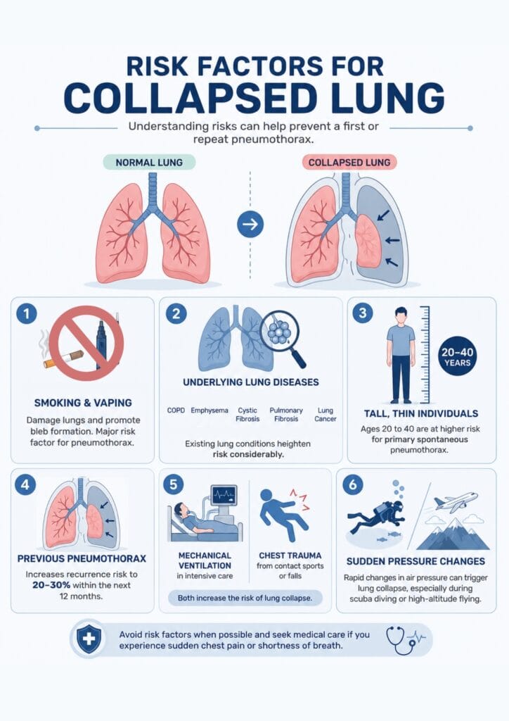
Understanding risk factors helps you make changes to reduce the likelihood of a first or repeat collapsed lung.
Major risk factors include:
Smoking (cigarettes, cigars) and vaping, which cause lung damage and promote bleb formation. Smoking is a major risk factor for developing a pneumothorax and increases the risk of lung collapse significantly.
Underlying lung diseases such as COPD, emphysema, cystic fibrosis, pulmonary fibrosis, and lung cancer. Existing lung conditions and underlying medical conditions heighten the risk considerably.
Being among tall, thin people aged 20 to 40 for primary spontaneous pneumothorax.
A previous pneumothorax, which increases recurrence risk to roughly 20 to 30 percent within the next 12 months.
Other contributors include mechanical ventilation in intensive care, chest trauma from contact sports or falls, genetic syndromes, and sudden pressure changes. Rapid changes in air pressure can trigger a lung collapse, especially during scuba diving or high-altitude flying.
Practical prevention:
Stop smoking and vaping. Seek structured cessation support.
After a pneumothorax, air travel is usually safe only after full radiological resolution, typically at least one to two weeks for simple cases. Individual advice from a specialist in respiratory medicine is essential.
Scuba diving is often permanently discouraged after a spontaneous pneumothorax unless definitive surgical repair has been performed and a diving physician has cleared you.
Specialist follow-up with a thoracic surgeon like Mr Marco Scarci is valuable to discuss recurrence risk, lifestyle modifications, and whether preventive surgery is advisable, and patients can arrange this through private thoracic surgery appointments in London.
"I had rib pain for 6 years, was bounced around NHS without diagnosis. I was recommended to Mr Scarci — at consultation within minutes he knew what the problem was. Was offered surgery date the next week. 2 weeks post surgery I am almost back to full health."
When to See a Thoracic Surgeon

Whilst many small pneumothoraces are managed by emergency and respiratory teams, recurrent or complex cases benefit from specialist thoracic surgical review. Referral is advisable when:
You have had multiple pneumothorax episodes on the same side or both lungs
A persistent air leak continues despite several days with a chest drain in place
Large bullae or blebs are visible on imaging, or underlying structural lung disease is suspected
Co-existing conditions such as emphysema, chest wall deformities or lung tumours are present
Mr Marco Scarci’s consultation includes thorough review of previous scans, chest drain records and operative notes, alongside discussion of symptoms, lifestyle needs and personal risk. Treatment options typically include VATS or robotic bullectomy to remove weakened areas of lung, and mechanical or chemical pleurodesis to adhere the lung to the chest wall and reduce recurrence risk. Aftercare includes follow-up appointments, pain management, return-to-work guidance and personalised advice on flying and diving.
Have a question about your diagnosis?
A consultation gives you a personalised plan — not a generic estimate.
FAQs
How long does recovery usually take?
A small spontaneous pneumothorax managed with observation may resolve within one to two weeks. Larger or post-surgical cases may need several weeks before full activity can resume. A follow-up chest X-ray confirms complete re-expansion before strenuous exercise or air travel is resumed.
Can a collapsed lung happen again after treatment?
Recurrence affects roughly 20 to 30% of patients within a year, particularly smokers and those with underlying lung disease. VATS bullectomy and pleurodesis significantly reduce the risk. Stopping smoking and avoiding unpressurised flights or deep diving without medical clearance are also important.
Is a collapsed lung always life-threatening?
Many small pneumothoraces are not immediately life-threatening but still require urgent medical assessment. Tension pneumothorax is different. It is a true emergency that can rapidly compromise the heart and the other lung. Any sudden chest pain combined with breathlessness should be treated as an emergency until serious causes have been excluded.
Can I fly or scuba dive after a lung collapse?
Flying is usually safe once the lung has fully re-expanded, typically at least one to two weeks after a simple case. Scuba diving is generally not recommended after a spontaneous pneumothorax unless definitive surgical repair and specialist diving clearance have been obtained.
Which specialist should I see if I think I am at high risk?
Sudden symptoms always warrant emergency department assessment first. Ongoing care is usually shared between a respiratory physician and a thoracic surgeon, particularly for recurrent cases or where keyhole surgery may prevent further episodes.
