

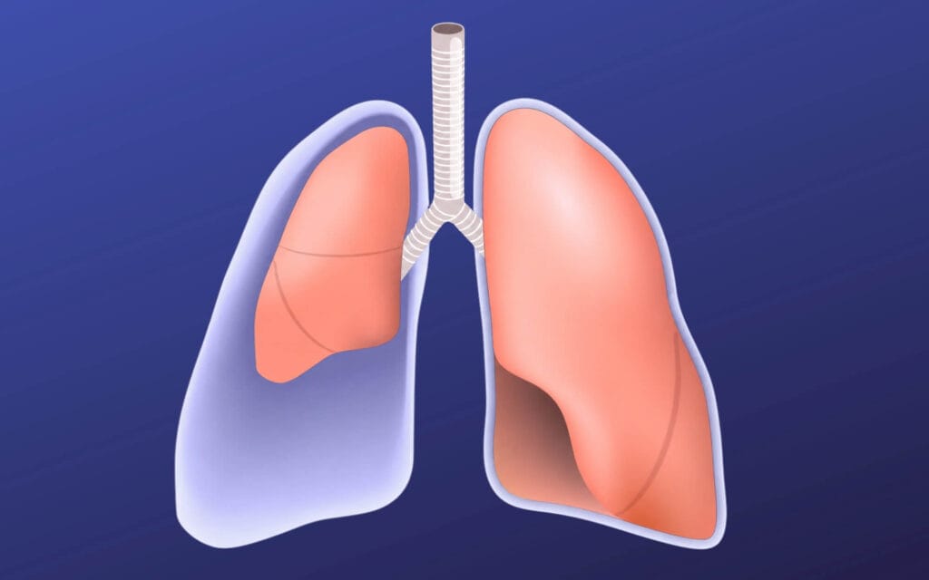
Collapsed lung pain catches most people off guard. It strikes suddenly, often without warning, and the experience can be alarming. Understanding where that pain occurs, what it feels like and when it signals a genuine emergency can help you act quickly and seek the right treatment.
Collapsed lung (pneumothorax) pain is typically sudden and sharp, felt on one side of the chest, often near the upper chest and shoulder, and may be accompanied by shortness of breath.
Pain usually worsens with deep breaths, coughing or movement and is often accompanied by chest tightness, rapid heart rate and difficulty breathing.
A large or tension pneumothorax is life-threatening and requires calling 999 immediately if severe chest pain and breathlessness develop together.
Only a physical examination and tests such as a chest X-ray or CT scan can confirm a collapsed lung. Chest pain alone is not enough to diagnose it.
What Is a Collapsed Lung (Pneumothorax)?
A pneumothorax occurs when air enters the pleural space (the thin gap between the lung surface and the chest wall), causing partial or complete lung collapse. This trapped air disrupts the normal pressure inside the pleural cavity, preventing the lung from expanding properly and resulting in reduced oxygen intake, impaired carbon dioxide removal and the sharp pain and breathlessness that define the condition.
Primary and secondary spontaneous pneumothorax are common in tall, thin young adults or in people with pre-existing lung conditions such as COPD or asthma. Traumatic pneumothorax follows a chest injury or medical procedure. The most dangerous form, tension pneumothorax, occurs when air continuously enters the pleural space but cannot escape, compressing the heart and the opposite lung, an immediately life-threatening emergency.
Mr Marco Scarci, a renowned consultant thoracic surgeon in London, sees pneumothorax as one of the most common reasons for urgent referral and specialist keyhole surgery, particularly when episodes recur.
Have a question about your diagnosis?
A consultation gives you a personalised plan — not a generic estimate.
Where Does Collapsed Lung Pain Occur in the Body?
Pain location depends on which lung is affected, the size of the collapse and whether surrounding structures such as the ribs, muscles or pleura are irritated.
Pain most commonly occurs on one side of the chest, typically the side of the affected lung, felt high in the chest near or behind the nipple line. Irritation of the diaphragm can also cause referred pain at the shoulder tip or neck via the phrenic nerve. Some patients feel pain in the upper back or under the shoulder blade, where pleural irritation extends posteriorly.
Patients often describe the sensation as “a knife” or “a pulled muscle” appearing suddenly at rest, whilst walking or after a cough. Pain felt between the shoulder blades can mislead people into thinking it is a muscle or spinal problem. Collapsed lung pain is usually localised to one side rather than spreading across the entire chest, though severe cases may feel more diffuse due to overall chest tightness and pressure.
"I had rib pain for 6 years, was bounced around NHS without diagnosis. I was recommended to Mr Scarci — at consultation within minutes he knew what the problem was. Was offered surgery date the next week. 2 weeks post surgery I am almost back to full health."
What Does a Collapsed Lung Feel Like?
Collapsed lung pain is usually sudden, sharp and directly linked to breathing, though intensity can range from mild discomfort to severe pain depending on the size of the collapse. It typically comes on within seconds, with a stabbing or tearing quality that worsens on taking a deep breath, coughing, laughing or bending. It sometimes eases slightly when sitting upright and staying still.
Alongside the pain, most patients experience shortness of breath or a feeling of being unable to take a full breath, accompanied by rapid shallow breathing, a fast heart rate and light-headedness. Chest tightness or a sense of pressure on the chest is also common.
A small pneumothorax may cause only a mild ache, a sense of catching in the chest or almost no symptoms at all, sometimes discovered incidentally on a chest X-ray. Research suggests up to 18% of patients delay seeking care for over a week when symptoms are mild. In tension pneumothorax, pain may be intense and accompanied by bluish lips or fingers, confusion, sweating and a feeling of impending doom, all of which require immediate emergency care.
Want a specialist opinion on your condition?
Get a clear answer from a surgeon who will be with you throughout.
When Collapsed Lung Pain Is an Emergency
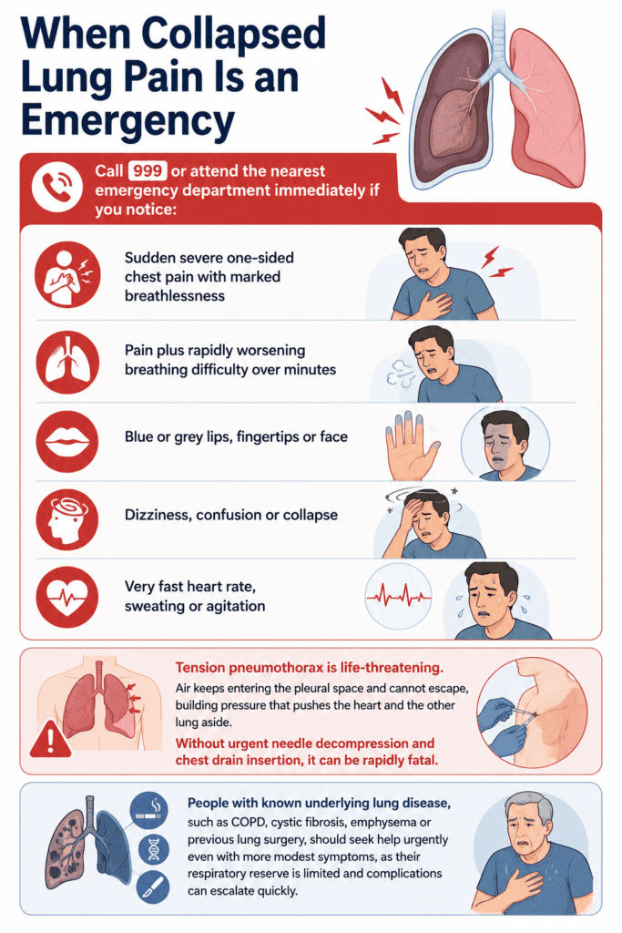
Call 999 or attend the nearest emergency department immediately if you notice:
Sudden severe one-sided chest pain with marked breathlessness
Pain plus rapidly worsening breathing difficulty over minutes
Blue or grey lips, fingertips or face
Dizziness, confusion or collapse
Very fast heart rate, sweating or agitation
Tension pneumothorax is life-threatening. Air keeps entering the pleural space and cannot escape, building pressure that pushes the heart and the other lung aside. Without urgent needle decompression and chest drain insertion, it can be rapidly fatal.
People with known underlying lung disease, such as COPD, cystic fibrosis, emphysema or previous lung surgery, should seek help urgently even with more modest symptoms, as their respiratory reserve is limited and complications can escalate quickly.
Causes and Risk Factors for Painful Lung Collapse
Collapsed lungs have several distinct causes. Primary spontaneous pneumothorax arises from the rupture of blebs on the lung surface in otherwise healthy lungs, most often in tall, thin adults aged 20 to 40. Secondary spontaneous pneumothorax occurs in people with underlying conditions such as COPD, emphysema, cystic fibrosis, pulmonary fibrosis or lung cancer, where already weakened lung tissue tears more easily. Traumatic pneumothorax is caused by physical damage to the chest: rib fractures, stab wounds, car accidents or medical procedures such as central line insertion or lung biopsy.
Several factors increase the likelihood of a collapsed lung. Cigarette smoking is among the most significant, as it promotes bleb disease and air leaks from damaged lung tissue. Men are more likely to experience pneumothorax than women, and a tall, thin body habitus is a well-recognised predisposing factor. A previous pneumothorax significantly increases the risk of recurrence, as do pre-existing lung conditions and certain genetic conditions such as Marfan syndrome.
These risk factors do not change the basic nature of the pain: sharp and pleuritic, but can influence how quickly symptoms progress and how often episodes recur. For those who recognise themselves in any of these categories, understanding what lies ahead is an important first step.
Not sure about your treatment options?
Mr Scarci provides expert consultations typically within one week of contact.
How Doctors Diagnose a Collapsed Lung
Many conditions, such as heart attack, blood clot, muscle strain and acid reflux, among them, can cause chest pain, so a structured medical assessment is essential.
Physical examination findings:
Reduced or absent breath sounds on one side
Increased chest wall movement on the opposite side
Reduced oxygen levels, raised pulse or low blood pressure
Tracheal deviation in severe cases
Main tests:
Test | Purpose |
Chest X-ray | Standard first test to detect air in the pleural space and estimate pneumothorax size |
CT scan | Detects small or complex pneumothoraces, blebs, bullae or underlying lung disease |
Bedside ultrasound | Rapid identification of lung collapse in emergency settings |
Treatment Options, Recovery and Long-Term Pain After a Collapsed Lung
Pneumothorax treatment depends on the size of the collapse, the severity of symptoms and whether the episode is a first or recurrent event. Small pneumothoraces may resolve on their own within days and are managed with observation and supplemental oxygen. Moderate cases are treated with needle aspiration, which removes trapped air through the chest wall. For larger or more symptomatic cases, a chest drain is inserted between the ribs to continuously remove air and allow the lung to re-expand.
Surgery is considered when pneumothorax recurs on the same side, when there is a bilateral collapse or persistent air leak, or in patients with high-risk occupations such as pilots or scuba diving professionals. Some patients also opt for surgery after a first severe episode. The most common surgical approach is video-assisted thoracoscopic surgery (VATS), which uses small keyhole incisions to remove fragile lung areas and adhere the lung to the chest wall. Compared with open surgery, VATS offers less post-operative pain, a shorter hospital stay and a quicker return to normal life.
Most patients recover within 2 to 4 weeks after VATS, though open surgery requires longer. Short-term pain around the incision sites and ribs is common and is usually managed with painkillers. Definitive surgical treatment significantly reduces the risk of recurrence, from roughly 300 per 1,000 patients down to 50 to 60 per 1,000 after surgery.
Have a question about your diagnosis?
A consultation gives you a personalised plan — not a generic estimate.
Frequently Asked Questions About Collapsed Lung Pain
Can a collapsed lung cause pain in the back or shoulder?
Yes. Pneumothorax pain can present as upper back or shoulder pain on one side, especially under the shoulder blade or at the shoulder tip, because the diaphragm and pleura share nerve pathways. Any new one-sided back or shoulder pain accompanied by breathlessness should be assessed urgently.
How long does pain last after treatment?
Acute sharp pain usually reduces significantly within a few days once the lung has re-expanded. A dull ache or chest wall soreness can persist for one to two weeks. After VATS surgery or chest drain insertion, local incision and rib pain may continue for several weeks but improves gradually. Persistent or worsening pain should be reviewed by your treating team.
Is it normal to feel anxious after a collapsed lung?
Many patients feel anxious, are reluctant to take deep breaths or worry about recurrence after a dramatic episode. This is entirely normal. Discussing these concerns openly with your thoracic surgeon or GP is encouraged, and referral for psychological support or breathing physiotherapy is available if needed.
Can I fly or scuba dive after a pneumothorax?
Flying is generally avoided until at least one week after full lung re-expansion has been confirmed on imaging, as changes in cabin air pressure can expand residual air and trigger pain or re-collapse. Scuba diving is usually discouraged permanently after spontaneous pneumothorax due to the high risk under pressure. Selected patients who have had definitive surgery may be individually assessed by a specialist.
Will lifestyle changes reduce the risk of another collapse?
Stopping smoking and vaping, avoiding recreational drugs, managing chronic lung conditions carefully and maintaining a healthy body weight can all reduce risk, though not eliminate it. Regular follow-up with a respiratory or thoracic specialist after any pneumothorax helps clarify your personal risk factors and whether preventive surgery would be appropriate.
