

Thoracic surgery has changed considerably over recent decades. Procedures that once required large incisions and rib spreading can now often be performed using keyhole or robotic-assisted techniques, supported by advanced imaging, refined instruments and structured recovery pathways.
These significant advancements have expanded the options available to patients with lung cancer, pneumothorax, pleural disease, mediastinal tumours and chest wall conditions. The most appropriate treatment still depends on the diagnosis, anatomy and general health of the individual, but many patients can now benefit from a more precise and less invasive approach.
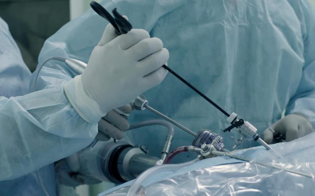
Minimally invasive surgery has become central to modern thoracic practice. VATS, robotic-assisted surgery and selected single-port techniques allow many procedures to be completed through smaller incisions without routine rib spreading.
For suitable patients, minimally invasive approaches can offer less post-operative pain, shorter hospital stays and faster recovery than traditional open thoracotomy.
Lung cancer surgery is increasingly coordinated with molecular testing, targeted therapy and perioperative immunotherapy, allowing treatment to be tailored more closely to the biology and stage of the tumour.
Advanced imaging, three-dimensional reconstruction, fluorescence guidance and improved surgical instruments support more accurate planning and dissection.
Enhanced Recovery After Surgery, prehabilitation, modern pain control and digital follow-up can help patients mobilise sooner and recover more confidently.
How Thoracic Surgery Has Changed Since the 1990s
In the 1980s and early 1990s, major thoracic surgery usually required a thoracotomy. This involved a long incision along the side of the chest, division of muscle and the use of a metal retractor to spread the ribs. Although it provided direct access to the chest cavity, patients often experienced considerable pain, prolonged hospital stays and a slow return to ordinary activities.
Modern thoracic surgery increasingly uses minimally invasive, precision-guided techniques. High-definition cameras, robotic systems, advanced imaging and specialised instruments allow surgeons to perform surgery through small incisions while aiming to achieve the same clinical or oncological goals as open surgery.
Video-assisted thoracoscopic surgery, commonly known as VATS, developed into a practical option for major lung procedures during the early 1990s. VATS lobectomy and wedge resection subsequently became established treatments for selected patients with early-stage lung cancer. Robotic-assisted thoracic surgery later introduced three-dimensional magnified vision and wristed instruments, while uniportal techniques reduced the number of incisions needed in suitable cases.
Lung cancer has remained an important driver of innovation because it is one of the leading causes of cancer death worldwide. However, the benefits extend beyond cancer. Minimally invasive techniques are also used for pneumothorax, emphysema, pleural infection, mediastinal tumours, hyperhidrosis and selected chest wall conditions.
Mr Marco Scarci is a consultant thoracic surgeon in London who uses VATS and robotic-assisted techniques in private and NHS practice. The choice of approach is based on the condition being treated, the patient’s anatomy and fitness, and whether a minimally invasive operation can achieve the safest and most effective result.
Have a question about your diagnosis?
A consultation gives you a personalised plan — not a generic estimate.
From Open Thoracotomy to Minimally Invasive Thoracic Surgery
Traditional open thoracotomy involves a long incision, muscle division and rib retraction. It remains an essential operation when direct access is required, but it can be associated with greater tissue trauma and a longer recovery.
By contrast, minimally invasive thoracic surgery uses small incisions, a camera to visualise the chest and specialised instruments to complete the procedure. There is usually no need for a metal rib spreader. For suitable patients, this can mean less pain, fewer wound complications and a shorter hospital stay.
Important stages in this transition include:
The development of modern VATS lung resection during the early 1990s;
Wider adoption of VATS lobectomy, wedge resection and pleural surgery during the 2000s;
Increasing use of robotic-assisted thoracic surgery across specialist UK and European centres;
Continued refinement of uniportal VATS and selected single-port robotic approaches.
Procedures such as lobectomy, segmentectomy, decortication, bullectomy, thymectomy and sympathectomy can now often be completed using minimally invasive techniques. Long-term surgical outcomes depend on complete tumour removal, appropriate lymph-node assessment, careful patient selection and the experience of the operating team rather than incision size alone.
UK thoracic practice has steadily moved towards minimally invasive lung resection. However, access and adoption vary between hospitals, and open surgery remains appropriate for very large tumours, complex central disease, extensive adhesions, major blood-vessel involvement and emergencies.
"After two years of being told my symptoms were anxiety, Mr Scarci identified the problem immediately. The surgery was straightforward and I was discharged after three days. I only wish I had found him sooner."
Video-Assisted Thoracoscopic Surgery (VATS): The Workhorse of Modern Thoracic Surgery
Video-assisted thoracoscopic surgery (VATS) uses a thoracoscope and specialised instruments inserted through small incisions between the ribs. The surgeon views the chest on a high-definition monitor and performs the operation without routinely spreading the ribs.
The number and size of incisions depend on the procedure and the surgeon’s technique. Conventional multi-port VATS often uses two to four access points, while uniportal VATS uses one incision in selected cases.
VATS is used for a wide range of procedures, including:
Lobectomy and segmentectomy for selected early-stage lung cancers
Wedge resection for small pulmonary nodules
Bullectomy and pleurodesis for recurrent pneumothorax
Decortication for pleural infection or empyema
Removal of selected mediastinal masses
Sympathectomy for severe hyperhidrosis
Evidence gathered over several decades supports VATS as an effective alternative to open thoracotomy for suitable patients. A systematic review may combine results from several studies to compare hospital stay, complications, pain scores and cancer outcomes, although differences in surgeon experience and patient selection must be considered.
Technical advances have made more complex VATS operations possible. These include high-definition and 4K cameras, angled thoracoscopes, powered stapling devices, improved vessel-sealing systems and more ergonomic instruments.
VATS is routinely considered for suitable patients with lung cancer, pleural disease, pneumothorax and benign nodules. Patient selection and pre-operative planning remain essential. A minimally invasive operation should only be chosen when it can provide safe access, complete treatment and an appropriate long-term outcome.
Want a specialist opinion on your condition?
Get a clear answer from a surgeon who will be with you throughout.
Robot-Assisted Thoracic Surgery (RATS): Precision Keyhole Surgery
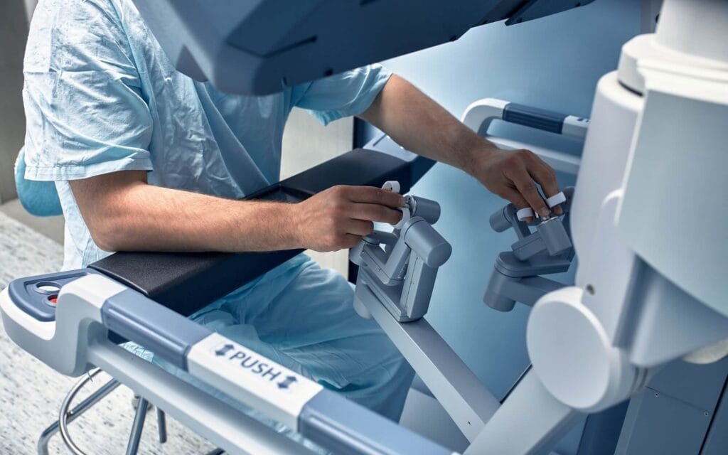
Robotic-assisted thoracic surgery (RATS) builds on the principles of keyhole surgery. The surgeon sits at a console in the operating theatre and controls robotic arms holding a camera and surgical instruments inside the patient’s chest.
The robotic system does not operate independently. Every movement is directed by the surgeon.
Potential technical advantages include:
A magnified three-dimensional view of the operating field
Wristed instruments with a greater range of movement than conventional VATS instruments
Tremor filtration
Improved ergonomics for the surgeon
Precise access to confined areas around the mediastinum, lymph nodes and central lung structures
Published comparisons generally suggest that VATS and robotic surgery provide broadly similar short-term results for many lung procedures. Robotic surgery may offer technical advantages during complex dissection and lymph-node assessment, although outcomes also depend on surgeon experience, case selection and the quality of the hospital programme.
Patients undergoing robotic lung cancer surgery may benefit from magnified three-dimensional vision and wristed instruments during complex dissection. However, robotic surgery should not automatically be considered superior for every patient.
Large surgical registries support quality improvement by tracking complications, conversion rates and other surgical outcomes across different healthcare systems. These datasets help teams compare performance while recognising that hospitals may treat patients with different levels of risk.
"I was diagnosed in March with a rare condition. My local hospital said it was inoperable due to the size of the mass. Mr Scarci reassured me the thymoma was resectable. I was booked for surgery the following week where he successfully removed the entire tumour."
Uniportal and Single-Port Techniques: Doing More Through One Incision
Uniportal VATS involves performing an operation through a single incision, commonly measuring approximately 3–4 cm. Uniportal robotic surgery follows a similar principle using a platform designed or adapted for single-port access.
The aim is to reduce the number of intercostal spaces used during surgery. Disturbing fewer spaces may reduce tissue trauma and post-operative discomfort, although pain is also influenced by incision position, nerve irritation, drain placement and the extent of the operation.
In experienced hands, uniportal VATS can be used for wedge resection, segmentectomy, lobectomy, pneumothorax surgery and selected complex procedures. Early findings suggest that uniportal robotic techniques may also be feasible, but the evidence base remains smaller than that for established multi-port VATS and robotic surgery.
Single-port surgery can present technical challenges, including instrument crowding and a demanding learning curve. Not every tumour or chest condition is suitable. Adhesions, central tumour location, vascular involvement and the need for complex reconstruction may favour a multi-port or open approach.
A specialist assessment can determine whether a single-port operation offers a genuine benefit without compromising safety or the completeness of treatment.
Not sure about your treatment options?
Mr Scarci provides expert consultations typically within one week of contact.
Advanced Techniques for Lung Cancer Surgery
Lung cancer, particularly non-small cell lung cancer, remains one of the most common indications for major thoracic surgery. Modern thoracic oncology increasingly tailors operations to the size, location and stage of the tumour, its molecular profile and the patient’s lung function.
The available lung cancer surgery options depend on the tumour’s location, stage and biological features, as well as the patient’s overall health.
Anatomical Segmentectomy
Anatomical segmentectomy removes one or more defined segments of a lung lobe rather than the entire lobe. It preserves more functioning lung tissue and may be appropriate for carefully selected small, peripheral tumours, including some cases of early lung adenocarcinoma.
Major trials have shown that sublobar resection can provide outcomes comparable to lobectomy in selected patients with small, node-negative peripheral non-small cell lung cancer.
These results do not mean that segmentectomy is suitable for every small tumour. Tumour characteristics, margins, lymph-node assessment and anatomy remain important. Study eligibility criteria may also be narrower than those encountered in routine clinical practice, so trial findings cannot automatically be applied to every patient.
Segmentectomy can be technically more complex than lobectomy because the surgeon must identify and divide individual segmental blood vessels and bronchi while preserving neighbouring lung tissue.
Sublobar Resection
Sublobar resection includes anatomical segmentectomy and wedge resection. A wedge resection removes the tumour with a margin of surrounding tissue without following the anatomical boundaries of a lung segment.
Modern VATS and robotic platforms support precise identification of vessels, bronchi and the boundaries between lung segments. Three-dimensional computed tomography reconstruction can help the surgeon understand individual anatomical variations before entering the operating theatre.
For selected central tumours, sleeve lobectomy or bronchoplastic and angioplastic procedures may remove an affected airway or blood-vessel segment while preserving more lung than a pneumonectomy. These highly specialised procedures may still require open surgery depending on their complexity.
Intraoperative Imaging
Frozen-section pathology allows tissue to be examined during surgery and may help confirm a diagnosis, assess a margin or evaluate a lymph node.
Fluorescence imaging may assist with identifying lung-segment boundaries, blood flow or selected nodules. Intraoperative ultrasound and localisation methods can also help find small or deep lesions that are difficult to feel during keyhole surgery.
These tools support accurate surgical resection but do not replace careful pre-operative staging or experienced surgical judgement. Different lung cancer operations may be considered according to tumour size, position and lung function.
Molecularly Targeted Therapy and EGFR-Positive Lung Cancer
What Is Targeted Therapy?
Targeted therapy uses medicines designed to interfere with particular molecular changes that help cancer cells grow. In non-small cell lung cancer, clinically important targets can include mutations in the epidermal growth factor receptor, known as EGFR, and rearrangements involving anaplastic lymphoma kinase, known as ALK.
These medicines are now an important part of lung cancer treatment, but they do not replace surgery for every patient. Molecular results are considered alongside cancer stage, lymph-node involvement, general health and the likelihood of complete removal.
Routine Molecular Testing
Molecular testing is an established part of lung cancer care, particularly when systemic treatment may be required. Testing can identify tumour changes that influence treatment after surgery or in advanced disease.
For resectable cancer, molecular findings are increasingly discussed by the multidisciplinary team before and after surgery. The precise tests required depend on tumour type, stage and national guidance.
Molecular information can also support risk stratification by identifying patients whose tumours may have a greater likelihood of recurrence or response to a targeted medicine. However, a risk-stratified classification should complement clinical staging rather than replace it.
EGFR Inhibitors and Surgical Planning
Third-generation EGFR inhibitors such as osimertinib have improved outcomes for eligible patients with resected EGFR-mutated non-small cell lung cancer when used as adjuvant treatment.
Research is also examining targeted therapy before surgery. Early studies suggest that neoadjuvant treatment may shrink some EGFR-mutated tumours, but this approach continues to evolve and is not identical to established post-operative treatment.
Treatment before surgery may alter the appearance and texture of tissues, occasionally making dissection more demanding. Thoracic surgeons and oncologists therefore coordinate treatment timing, the planned extent of resection and lymph-node assessment.
After surgery, follow-up usually involves clinical review and imaging. Circulating tumour DNA is being studied as a form of liquid biopsy that may help detect microscopic residual disease, but it has not replaced standard imaging surveillance.
Have a question about your diagnosis?
A consultation gives you a personalised plan — not a generic estimate.
Perioperative Immunotherapy: Harnessing the Immune System Around Surgery
What Is Perioperative Immunotherapy?
Perioperative immunotherapy refers to immune checkpoint inhibitors given before surgery, after surgery or during both periods. These medicines help the immune system recognise and attack cancer cells.
Drugs such as nivolumab and pembrolizumab are now used in selected patients with resectable non-small cell lung cancer, often alongside chemotherapy. Eligibility depends on cancer stage, molecular profile, general health and national treatment guidance.
Key Clinical Trial Findings
The CheckMate 816 trial established the benefit of nivolumab plus chemotherapy before surgery for selected patients with resectable non-small cell lung cancer.
At five years, overall survival was 65.4% in the intervention group receiving nivolumab plus chemotherapy, compared with 55.0% in the chemotherapy-alone group. The findings suggest that combining immunotherapy with chemotherapy before surgery can improve outcomes for appropriately selected patients.
The original trial also found a pathological complete response in 24.0% of patients receiving nivolumab plus chemotherapy, compared with 2.2% receiving chemotherapy alone.
A major pathological response generally means that 10% or less viable tumour remains in the surgical specimen following pre-operative treatment. A pathological complete response means that no viable tumour is identified.
Practical Surgical Implications
Pre-operative immunotherapy and chemotherapy may shrink a tumour or reduce cancer in the lymph nodes. This can improve the likelihood of complete removal in some patients.
Treatment can also cause fibrosis, inflammation or thickening around the tumour and lymph nodes, potentially making dissection more difficult. Experienced surgeons can still perform many of these operations through VATS or robotic techniques, but conversion to open surgery may be required when this is the safest option.
Mr Scarci works within a multidisciplinary setting so that imaging, systemic treatment, surgery, pathology and follow-up are coordinated. This allows the sequence of treatment to reflect the individual tumour rather than relying on a single standard pathway.
Oesophageal Cancer and Complex Mediastinal Surgery
Oesophageal cancer has also benefited from minimally invasive and robotic-assisted techniques, particularly for tumours affecting the thoracic oesophagus.
A minimally invasive oesophagectomy may combine laparoscopic or robotic mobilisation of the stomach with thoracoscopic or robotic mobilisation of the oesophagus. The stomach is fashioned into a tube and joined to the remaining oesophagus in the chest or neck.
Compared with open oesophagectomy, minimally invasive surgery may reduce pulmonary complications and support faster recovery in suitable patients. Outcomes depend heavily on careful selection, multidisciplinary treatment and the experience of the surgical centre.
Chemotherapy, radiotherapy and immunotherapy may form part of treatment depending on tumour type and stage. Accurate staging commonly includes CT, PET-CT, endoscopy, biopsy and sometimes endoscopic ultrasound.
Patients with gastro-oesophageal reflux disease may also undergo assessment of the oesophagus and hiatus when symptoms, imaging or the planned operation indicate that this is relevant. However, reflux disease is distinct from oesophageal cancer and does not normally require thoracic cancer surgery.
Mediastinal surgery has undergone a similar transformation. Thymomas and other selected anterior mediastinal masses that once routinely required a sternotomy can now sometimes be removed through VATS or robotic-assisted surgery.
The safest approach depends on tumour size, invasion into nearby structures and the need for complete removal. Open surgery remains appropriate when a mass involves major blood vessels or other critical structures.
Want a specialist opinion on your condition?
Get a clear answer from a surgeon who will be with you throughout.
Thoracic Surgery for Benign Conditions: Pneumothorax, Emphysema and Hyperhidrosis
Not all thoracic surgery is performed for cancer. Keyhole surgery is also used for several benign but potentially debilitating conditions.
These include recurrent pneumothorax, advanced emphysema, severe hyperhidrosis, pleural infection, chest wall deformities and selected rib problems. They form part of the wider range of thoracic conditions treated by specialist thoracic teams.
Modern VATS treatment for recurrent primary spontaneous pneumothorax commonly involves removing visible blebs or bullae and performing a procedure that encourages the lung to adhere to the chest wall. Depending on the patient and procedure, hospital discharge may be possible within a few days.
Patients undergoing pneumothorax surgery are assessed according to the cause of the collapse, recurrence risk, imaging and overall health. The operation can reduce recurrence risk, although no treatment can guarantee that another collapse will never occur.
For selected patients with severe emphysema, lung-volume-reduction surgery removes the most damaged areas so that the remaining lung and breathing muscles can work more effectively. Assessment may include the CT distribution of emphysema, exercise capacity and forced expiratory volume, usually abbreviated as FEV1.
Endoscopic thoracic sympathectomy is a minimally invasive procedure for severe palmar or axillary hyperhidrosis. It interrupts part of the sympathetic nerve chain through small incisions. The procedure can be highly effective, but patients need a detailed discussion about possible adverse effects, particularly compensatory sweating.
Chest Wall and Rib Surgery: 3D Planning and Reconstruction
Chest Wall Tumour Resection and Reconstruction
Modern management of chest wall tumours may require removal of the lesion together with a margin of surrounding tissue. When a significant section of the chest wall is removed, reconstruction may be required to protect the internal organs and preserve breathing mechanics.
Reconstruction options include titanium plates, surgical mesh and custom-designed implants. Three-dimensional modelling based on CT imaging can help plan complex resections and, in selected cases, support the design of patient-specific prostheses.
These techniques aim to restore stability while achieving appropriate tumour clearance. VATS or robotic surgery may sometimes be combined with an open chest wall resection when underlying lung or pleural tissue must also be removed.
Structural chest problems such as chest wall deformities require assessment of symptoms, breathing, appearance and their effect on everyday activity.
Rib Fracture Stabilisation
Most rib fractures heal without surgery. However, surgical stabilisation may be considered for selected patients with flail chest, multiple severely displaced fractures, chest wall instability, respiratory compromise or painful non-union.
The procedure uses plates and screws to stabilise the fractured ribs. Pre-operative CT imaging helps map the fracture pattern and plan the most appropriate access.
Three-dimensional reconstruction may assist with complex injuries, but decisions are based on breathing, pain, displacement, associated injuries and general health rather than imaging alone.
Not sure about your treatment options?
Mr Scarci provides expert consultations typically within one week of contact.
Imaging, Navigation and 3D Reconstruction in Thoracic Surgery
Pre-operative Planning With 3D Reconstruction
Modern CT and PET-CT imaging allow detailed mapping of tumours, blood vessels, airways and lymph nodes before surgery.
Three-dimensional reconstruction software converts computed tomography data into virtual models of the patient’s lungs and chest. These models may reveal anatomical variations and help the surgeon plan a segmentectomy or locate a small pulmonary nodule.
This strengthens pre-operative planning, particularly in minimally invasive surgery where the surgeon cannot always feel the entire lung directly. However, three-dimensional planning remains an aid rather than a substitute for anatomical knowledge and intraoperative judgement.
Pulmonary Nodule Malignancy Assessment
Assessment of pulmonary nodule malignancy considers the size, shape, growth rate and density of a nodule, together with the patient’s age, smoking history and previous cancer history.
Risk-stratification models can help estimate the likelihood that a nodule is malignant. A risk-stratified classification may then guide whether the patient needs CT surveillance, PET-CT, biopsy or surgical assessment.
These models support clinical decision-making but do not provide certainty. Some cancers appear low-risk on imaging, while many benign nodules have features that initially appear suspicious.
Augmented Reality and Navigation
Augmented reality and mixed-reality systems are being investigated as ways of placing virtual anatomical information within the surgeon’s field of view.
Early findings suggest that these technologies may support localisation of small nodules and improve spatial understanding during complex procedures. However, widespread use remains limited, and accuracy, workflow, cost and clinical benefit require continued evaluation.
Intraoperative Localisation Tools
Very small or deep lung nodules may be difficult to identify during VATS or robotic surgery. Localisation options include electromagnetic or robotic bronchoscopy, fluorescent markers, dyes, wires and microcoils placed before or during surgery.
Single-modality imaging may not always provide all the information required. Fusing CT, PET-CT, bronchoscopy and three-dimensional reconstruction can give the surgical team a more complete anatomical picture.
These approaches aim to help the surgeon remove a lesion accurately while preserving as much healthy lung tissue as possible.
Artificial Intelligence and Decision Support in Thoracic Surgery
Artificial intelligence is being explored across thoracic care, including lung-cancer screening, pulmonary-nodule assessment, surgical planning, risk prediction and post-operative monitoring.
AI systems can analyse CT scans and highlight areas that may require closer review. They may also help estimate pulmonary nodule malignancy and support risk stratification when large numbers of scans need assessment.
However, sensitivity and accuracy vary between systems, datasets and patient populations. An AI result cannot confirm or exclude cancer by itself. Clinical history, comparison with previous scans, specialist radiology review and, where appropriate, biopsy remain essential.
AI-supported three-dimensional reconstruction can speed up the separation of lung segments, vessels and bronchi on CT imaging. Predictive models are also being studied for potential clinical applications such as estimating post-operative risk and response to neoadjuvant therapy.
Why Prospective Multicentre Validation Matters
Most current AI systems are decision-support tools rather than replacements for clinical judgement. Before an emerging system is adopted widely, prospective multicentre validation is needed to confirm that it performs reliably across different hospitals, scanners, clinical teams and patient populations.
Results from one specialist centre may not transfer directly to routine care elsewhere. Wider prospective validation and external validation can identify limitations, variation in accuracy and groups for whom a system may be less reliable.
Research reviews should explain clearly how evidence was identified, assessed and compared. Patient-facing information should then translate those findings into practical language rather than reproducing technical academic reporting.
Study Eligibility and Patient Selection
Study eligibility criteria and patient selection are particularly important when AI tools or new surgical techniques are evaluated.
A system tested only on carefully selected scans may not perform as well in people with previous surgery, unusual anatomy, severe lung disease or lower-quality imaging. Researchers and clinicians must therefore assess whether the people included in a study reflect the patients seen in everyday practice.
The final recommendation must reflect the diagnosis, anatomy, health and preferences of the patient, even when an AI model provides a risk-stratified classification.
Have a question about your diagnosis?
A consultation gives you a personalised plan — not a generic estimate.
Instruments, Staplers and Energy Devices: Quiet Revolutions at the Operating Table
Many advances in thoracic surgery come from instruments that receive less attention than robotic systems but have changed what can be achieved through keyhole incisions.
Modern endoscopic staplers allow surgeons to divide pulmonary arteries, veins, bronchi and lung tissue through small access ports. Powered firing and controlled tissue compression may improve consistency, although safe use still depends on correct positioning and tissue assessment.
Different staple heights are available for tissues of varying thickness. This can help achieve a secure closure while reducing unnecessary compression.
Ultrasonic and bipolar energy devices allow controlled cutting and vessel sealing during lung and mediastinal dissection. They can reduce the need for clips or sutures in selected tissues and help maintain a clear operating field.
High-definition cameras, three-dimensional imaging and fluorescence systems improve visualisation of anatomical structures, blood flow and tissue boundaries.
When combined with surgeon experience and careful planning, these instruments allow increasingly complex cancer and benign operations to be performed through smaller incisions.
Perioperative Care, Anaesthesia and Airway Management Innovations
Many thoracic procedures require one-lung ventilation, during which one lung is allowed to collapse while the other is ventilated. This provides space for the surgeon to work inside the chest.
Anaesthetic techniques and ventilation strategies have become more refined, with greater emphasis on protecting both lungs and maintaining stable oxygen levels.
Modern approaches may include:
Lower tidal volumes and individually adjusted positive end-expiratory pressure
Careful fluid management to reduce the risk of lung oedema
Bronchodilator or inhaler optimisation for patients with asthma or COPD
Advanced monitoring of oxygenation, ventilation and cardiovascular function
Regional anaesthetic techniques to reduce post-operative opioid requirements
Non-intubated thoracic surgery is performed in selected specialist settings. It uses sedation and regional anaesthesia rather than conventional general anaesthesia with a breathing tube.
Although the approach may reduce some airway-related effects, it is not suitable for every operation or patient and requires an experienced surgical and anaesthetic team.
Modern anaesthesia is an essential partner to minimally invasive surgery. Effective ventilation, pain relief and monitoring help patients breathe comfortably and begin moving soon after their operation.
Want a specialist opinion on your condition?
Get a clear answer from a surgeon who will be with you throughout.
Enhanced Recovery After Surgery (ERAS) in Thoracic Practice
Enhanced Recovery After Surgery (ERAS) is a structured approach covering preparation, anaesthesia, pain control, mobilisation, nutrition and discharge planning.
Rather than relying on one intervention, ERAS combines multiple evidence-based steps designed to reduce the physical stress of surgery and support a safer recovery.
Before Surgery
Smoking cessation, nutritional assessment, prehabilitation, medication review and clear patient counselling can improve readiness for surgery. Prolonged fasting is avoided where possible, following instructions from the anaesthetic team.
During Surgery
Minimally invasive access, lung-protective ventilation, multimodal pain relief and careful fluid management help limit surgical stress.
Post-operative Management
Modern post-operative management encourages patients to sit out of bed and mobilise early. Chest drains and urinary catheters are removed when clinically appropriate, while oral food and fluids are restarted promptly.
Pain, breathing, oxygen levels, mobility and wound healing are monitored throughout the hospital stay. Discharge planning begins early so that patients understand medication, activity, wound care and the warning signs that require medical advice.
ERAS programmes are associated with shorter hospital stays and fewer complications after lung surgery when implemented consistently. They also provide clearer daily recovery goals.
Mr Scarci incorporates ERAS principles into his NHS and private pathways so that preparation, surgery and follow-up form part of one coordinated recovery plan.
Evidence From a Multicentre Cohort
Evidence from a multicentre cohort can indicate whether an ERAS pathway produces consistent benefits across hospitals with different teams, resources and patient populations.
Evidence across several centres is often more representative of routine practice than results from one specialist hospital. However, differences in patient selection, surgical techniques and discharge policies must still be considered when interpreting outcomes.
Prehabilitation and Pulmonary Rehabilitation Before and After Thoracic Surgery
Prehabilitation involves targeted exercise, breathing training, nutritional support and psychological preparation before surgery.
It may be particularly helpful for patients with reduced fitness, COPD, poor nutritional status or limited lung function. Even a relatively short programme can improve confidence and functional capacity before an operation.
A typical programme may include:
Supervised aerobic and resistance exercise
Inspiratory muscle training
Smoking-cessation support
Dietary advice and protein optimisation
Education about breathing exercises and recovery
Psychological support where needed
Evidence suggests that pre-operative exercise can reduce pulmonary complications in selected lung-surgery patients, although the size of the benefit varies between programmes and patient groups.
After surgery, physiotherapy, progressive walking and appropriate pulmonary rehabilitation can help patients regain strength and confidence. Measurements such as forced expiratory volume may be used to assess respiratory function before and after treatment.
Smoking cessation, nutrition, rest and gradual activity all support healing after surgery, although recovery recommendations should be tailored to the operation and the individual patient.
Not sure about your treatment options?
Mr Scarci provides expert consultations typically within one week of contact.
Preventing a Post-operative Pulmonary Complication
A post-operative pulmonary complication is a breathing-related problem that develops after surgery. Examples include pneumonia, partial lung collapse, retained mucus, respiratory failure or a prolonged need for oxygen support.
The risk is influenced by the type of operation, existing lung disease, smoking, age, fitness, pain control and the ability to cough and mobilise.
Prehabilitation can reduce risk before surgery, while effective pain relief, breathing exercises, supported coughing and early movement remain important afterwards. Patients with COPD or asthma may also need their inhaler treatment reviewed before admission.
Minimally invasive surgery can reduce tissue trauma, but it does not remove the possibility of respiratory complications. Close monitoring and early treatment remain essential for most patients undergoing major lung surgery.
Modern Approaches to Pain Control and Analgesia
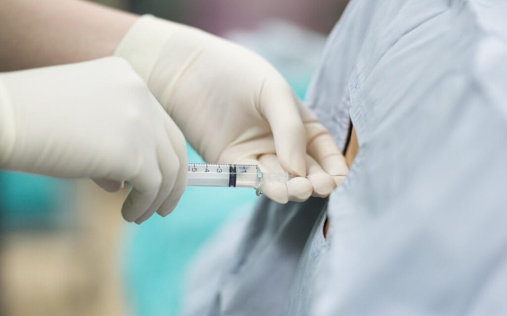
Effective pain control after thoracic surgery is essential because pain can limit deep breathing, coughing and movement.
Poorly controlled pain may increase the risk of mucus retention, chest infection, blood clots and delayed recovery. The aim is to provide enough relief for patients to breathe and mobilise while limiting medication-related side effects.
Regional techniques include:
Paravertebral blocks, in which local anaesthetic is placed beside the spine
Erector spinae plane blocks, targeting tissue planes over the muscles beside the spine
Serratus anterior plane blocks, which can numb the side of the chest
Local wound infiltration, placing anaesthetic around the incision
Thoracic epidural analgesia, which remains appropriate for selected major open procedures
The best option depends on the procedure, incision, health of the patient and anaesthetic assessment. Studies may compare opioid use and pain scores after different techniques, but results are influenced by the type of surgery and the broader pain-relief programme.
Multimodal analgesia combines medicines that work in different ways. These may include paracetamol, anti-inflammatory medication where safe, local anaesthetics and limited opioids.
Gabapentinoids are not suitable for every patient and may cause sedation, dizziness or other adverse effects. Their use should be individualised rather than routine.
Have a question about your diagnosis?
A consultation gives you a personalised plan — not a generic estimate.
Digital Health, Wearables and Remote Follow-up
Smartphones, wearable devices and secure telemedicine platforms are increasingly being used to support recovery after thoracic surgery.
Electronic patient-reported outcome systems allow patients to record symptoms such as pain, breathlessness, cough, wound concerns and mood. This may help teams identify deterioration earlier, although these systems do not replace emergency assessment or direct clinical review.
Wearable devices can record steps, heart rate, sleep and, in some cases, oxygen saturation. Changes may help clinicians understand recovery trends, but consumer devices vary in accuracy and should not be used alone to diagnose complications.
Virtual consultations can help patients discuss scan results, wound healing and recovery without travelling to London for every appointment. They are particularly useful for people living elsewhere in the UK or overseas.
A virtual consultation can be used to review symptoms, available imaging and possible next steps when an in-person examination is not initially required.
Digital follow-up is not suitable for every concern. New breathlessness, chest pain, fever, significant wound changes or other urgent symptoms may require an in-person examination or emergency assessment.
What These Advances Mean for Patients Choosing Thoracic Surgery in London
VATS, robotic surgery, improved imaging, modern systemic therapy, ERAS and digital follow-up can provide meaningful benefits for selected patients.
These may include:
Smaller incisions and less visible scarring
Reduced post-operative pain
Earlier mobilisation
Shorter hospital stays
More precise anatomical planning
Better coordination between surgery and oncology
Improved support before and after discharge
A modern patient pathway may include:
Initial Consultation
The first appointment may be face-to-face or virtual. Symptoms, previous treatment, CT or PET-CT imaging, biopsy results and lung-function tests are reviewed.
Multidisciplinary Discussion
For cancer, the case is considered by surgeons, oncologists, radiologists, respiratory physicians, pathologists and other specialists. The team assesses whether surgery is appropriate and whether treatment should be given before or after the operation.
Pre-operative Planning
Pre-operative planning may involve detailed CT assessment, molecular testing, cardiac or respiratory review and prehabilitation for patients who would benefit from improved fitness before surgery.
The surgical approach is selected according to the diagnosis, anatomy and expected complexity. VATS or robotic surgery may be preferred when it can provide complete treatment safely, while open surgery remains important for selected advanced or technically complex cases.
Surgery
The procedure may use VATS, robotic-assisted surgery or an open approach. The safest method is the one that allows complete and effective treatment for the individual patient.
Structured Follow-up
Follow-up may include wound review, pathology discussion, imaging surveillance, rehabilitation and coordination with oncology or respiratory teams.
Both private and NHS pathways in London can provide access to modern thoracic care, although waiting times, hospital choice and local availability of robotic technology may differ. The options for private thoracic surgery depend on the diagnosis, chosen hospital, insurance and clinical requirements.
Many patients with lung, pleural or chest wall conditions can now avoid a traditional thoracotomy. However, open surgery remains necessary when it provides safer access or a better chance of complete treatment.
Want a specialist opinion on your condition?
Get a clear answer from a surgeon who will be with you throughout.
Frequently Asked Questions
How Long Will I Stay in Hospital After Minimally Invasive Lung Surgery?
Most patients having VATS or robotic lobectomy stay around 3–5 days, while smaller procedures may only require 1–3 days. Discharge depends on stable breathing, controlled pain, safe mobility and appropriate chest-drain management. The team also monitors for issues such as pneumonia, mucus retention or incomplete lung re-expansion.
Am I a Candidate for Minimally Invasive VATS or Robotic Surgery?
Candidacy is based on the condition being treated, tumour characteristics, anatomy, previous treatment or surgery, and overall fitness, not age alone. CT scans and lung-function testing help plan the safest approach. Previous surgery, radiotherapy or complex disease can make keyhole surgery harder, but do not automatically rule it out.
Is Thoracic Surgery Safe for Older Patients or Those With COPD or Heart Disease?
Many older patients and those with well-managed lung or heart conditions can have thoracic surgery safely following thorough assessment. Testing may include lung-function checks, heart tests and exercise assessment. Minimally invasive techniques can support faster recovery, but risk is personalised based on factors such as frailty, lung capacity, heart function and the operation required.
What Is the Difference Between Thoracic Surgery Privately and on the NHS in London?
Both NHS and reputable private hospitals should follow the same principles of safe, evidence-based care and multidisciplinary review. Private care can provide faster appointments, more flexibility and hospital choice, while the NHS offers comprehensive treatment, particularly for urgent or complex cancer cases. Some patients use a combination of private and NHS care.
How Can I Prepare for the Best Possible Outcome?
Stop smoking early, stay as active as your health allows, and follow any breathing or exercise advice. Take medication as directed, follow fasting and admission instructions, and organise help at home for recovery. Prehabilitation, physiotherapy, nutrition or psychological support may also help improve fitness and confidence before surgery.
