

Minimally invasive thoracic surgery has transformed the way surgeons treat conditions of the lungs, chest wall, and mediastinum. Instead of a large open cut that spreads the ribs apart, operations are now routinely performed through small keyhole incisions using cameras and specialised instruments. This article explains what minimally invasive thoracic surgery involves, how it works in practice, who it can help, and what patients should expect before, during, and after the procedure.
Minimally invasive thoracic surgery uses small keyhole incisions, typically 2–4 cuts of just 1–3 cm each, a camera, and specialised instruments instead of a large incision and rib spreading.
The two main approaches are video-assisted thoracoscopic surgery (VATS) and robotic-assisted thoracic surgery. Both approaches can reduce pain, scarring, hospital stay, and recovery time compared with open thoracotomy.
In Mr Marco Scarci’s London thoracic surgery practice, minimally invasive approaches are the default for suitable patients. Minimally invasive techniques are commonly used for lung cancer, pneumothorax, mediastinal tumours, and other chest conditions.
Open surgery may still be safer for very large tumours, complex disease involving major blood vessels, or emergencies.
Many patients can be assessed for a minimally invasive approach, but suitability depends on the condition, anatomy, and overall health.
Understanding Minimally Invasive Thoracic Surgery
Thoracic surgery covers operations on the lungs, the pleura (the thin membrane lining the lung and chest cavity), the mediastinum (the central compartment between the lungs containing the thymus, major blood vessels and other structures) and the chest wall, including the ribs. Traditionally, these operations were performed through a large open thoracotomy, involving a long incision down the side of the chest, cutting through muscles and using heavy retractors to spread the ribs apart so the surgeon could see and work inside.
Minimally invasive thoracic surgery replaces that large opening with several small incisions, typically 1–3 cm each, placed between the ribs. The term covers two main techniques: video-assisted thoracic surgery (VATS) and robotic-assisted thoracic surgery. In both, the surgeon operates using a high-definition camera called a thoracoscope, along with long, fine instruments passed through keyhole ports. Every movement is controlled by the surgeon and, despite the name, the surgical robot does not act independently.
These minimally invasive techniques aim to minimise tissue trauma while achieving the same surgical goals as open surgery, including removing diseased lung tissue, sampling lymph nodes, draining infections and excising tumours. Common procedures include lung surgery for cancer or benign nodules, pleural procedures, removal of mediastinal tumours such as thymoma and chest wall operations. The minimally invasive approach is a core part of modern cardiothoracic surgery and plays a central role in lung cancer surgery decision-making and techniques.
Not sure about your treatment options?
Mr Scarci provides expert consultations typically within one week of contact.
How Does Minimally Invasive Thoracic Surgery Work in Practice?
From the patient’s perspective, a minimally invasive thoracic operation begins much like any other surgery. You arrive in the operating room, meet the anaesthetic team, and receive general anaesthesia. A breathing tube is placed to ventilate one lung while the other is deflated, giving the surgeon space to work inside the thoracic cavity.
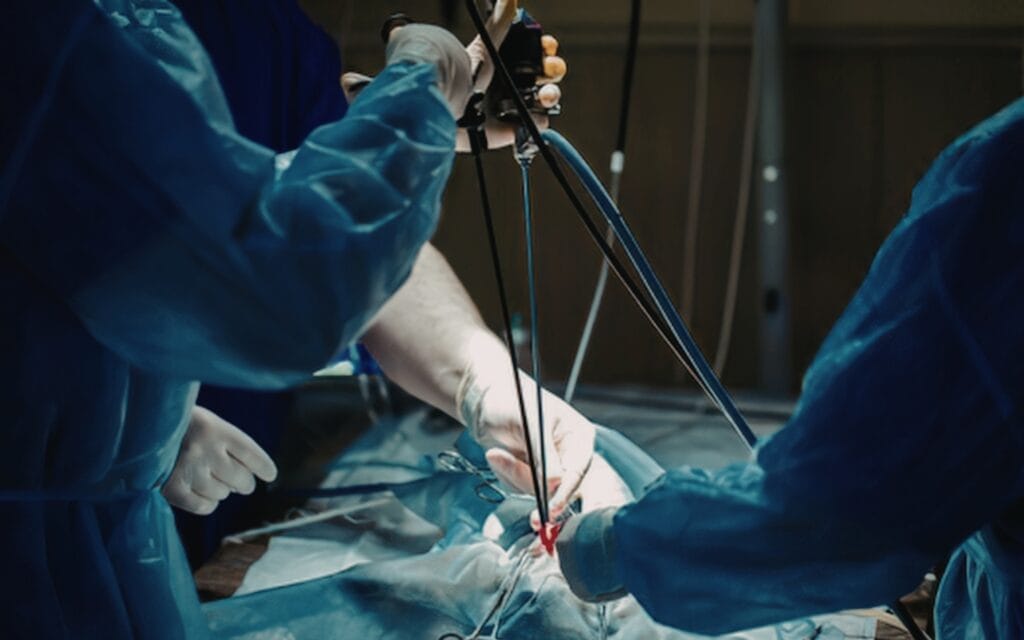
You are then positioned on your side with the arm raised. The surgeon makes 2–4 small incisions between the ribs. Minimally invasive thoracic surgery uses small incisions for access, typically just 1–3 cm. One port receives the thoracoscope, which sends magnified high-definition video to screens in the operating room. The remaining ports accept specialised instruments for cutting, grasping, and sealing tissues.
Crucially, there is no metal retractor spreading the ribs apart. The surgeon watches the screen and uses the instruments to carefully dissect around blood vessels, bronchi, and lymph nodes. Diseased lung tissue or tumours are freed from surrounding structures, placed in a protective surgical bag, and extracted through one of the small incisions, sometimes with a slight extension if the specimen is larger. Tissue samples are sent for analysis.
At the end of the operation, one or two chest drains (a chest tube) are placed in the pleural space to remove residual air and fluid. The small incisions are closed with dissolvable stitches or fine sutures.
How does this compare with open thoracotomy? In traditional thoracotomy, a single cut of 15–20 cm is made, major chest muscles are divided, and a retractor cranks the ribs apart, sometimes fracturing one. The result is significantly more surgical trauma, more post-operative pain, and a longer recovery. VATS procedures typically involve three or four small incisions, and the tissue damage is a fraction of what occurs with larger incisions.
"Dr. Marco Scarci is a deeply knowledgeable, open-minded, and empathetic surgeon. He listens to details and solves problems to truly deliver the most optimal results. As a medical professional myself, I can tell you that finding a specialist like Dr. Scarci is very rare, and it is a privilege to have him in my corner."
Video-Assisted Thoracoscopic Surgery (VATS): The Classic Keyhole Approach

Video-assisted thoracoscopic surgery (VATS) is the original and most widely used form of minimally invasive thoracic surgery. The first successful VATS lobectomy was performed in 1991, and the technique has since become routine worldwide. By 2015, 70% of lung operations at Stanford used VATS techniques, reflecting the global shift towards keyhole approaches in thoracic surgery.
A standard VATS set-up uses 2–4 ports. The incisions are typically 1–2 cm long, with one used for the camera and the others for surgical instruments. A more advanced development, known as uniportal VATS, uses a single incision to minimise surgical trauma. It may be used in selected cases, including early-stage lung cancer surgery and pneumothorax repair.
VATS lobectomy has become the gold standard for many cases of early-stage non-small cell lung cancer. It provides cancer control comparable to open surgery while offering less pain and a quicker recovery.
Common VATS procedures include:
Lung biopsy and wedge resection to diagnose or remove small nodules
Lobectomy and segmentectomy for lung cancer
- Pleurodesis for recurrent pneumothorax, often as part of comprehensive pneumothorax treatment by a specialist thoracic surgeon
Decortication for pleural infection, also known as empyema
Lymph node dissection for cancer staging
The technique provides excellent visualisation of the chest cavity, less muscle disruption, reduced blood loss and shorter hospital stays. However, VATS requires advanced surgical training. Fellowship programmes provide structured training in minimally invasive techniques, and surgeons may need to complete 30–50 VATS cases to achieve proficiency. Simulation-based training also plays an important role in developing the skills required for minimally invasive surgery.
The effectiveness of minimally invasive thoracic surgery is closely linked to the surgeon’s experience. Mr Marco Scarci, a specialist thoracic surgeon in London, has extensive experience in VATS techniques gained across high-volume centres and now applies this expertise in both his private and NHS practice.
Want a specialist opinion on your condition?
Get a clear answer from a surgeon who will be with you throughout.
Robotic-Assisted Thoracic Surgery: How Robotic Surgery Enhances Precision
Robotic-assisted surgery uses platforms such as the da Vinci Surgical System, the leading surgical robot in thoracic surgery, to give the surgeon enhanced control during complex procedures. Robotic-assisted surgery was first cleared by the FDA in 2000, and its use in thoracic surgery has grown steadily since. The surgeon sits at a console in the operating room, controlling robotic arms that enter the patient’s chest through keyhole incisions similar in size to VATS ports.
The main technical advantages of robotic surgery are significant:
3D high-definition visualisation: Robotic-assisted surgery offers enhanced 3D high-definition visualisation with up to 10× magnification, providing a depth of view that is not possible with standard VATS cameras.
Wristed instruments with seven degrees of freedom: This is greater than the human hand and enables enhanced dexterity and precise movements in confined spaces.
Tremor filtration: The system filters the natural slight movement of the surgeon’s hands, supporting greater precision during delicate dissection.
Robotic thoracic surgery can be particularly beneficial for complex procedures, including centrally located lung cancers near major blood vessels, lobectomy and segmentectomy requiring careful lymph node dissection, resection of mediastinal tumours such as thymoma, and selected chest wall operations. The da Vinci system’s 3D high-definition visualisation is especially valuable when operating in tight or anatomically challenging areas.
Compared with VATS, robotic-assisted lung cancer surgery can involve slightly longer operating times during the early learning curve. Robotic surgery training may require 20–80 cases to achieve proficiency. However, the platform may offer superior dexterity in confined spaces and an improved lymph node harvest. One study found a median of five lymph node stations sampled with robotic surgery compared with three using VATS. RATS may also lead to shorter hospital stays than open surgery, with published comparisons reporting approximately 6.9 days versus 8.0 days.
For suitable patients, Mr Marco Scarci collaborates with centres equipped with robotic platforms, recommending robotic-assisted surgery when its added precision may provide a clear clinical benefit.
"After two years of being told my symptoms were anxiety, Mr Scarci identified the problem immediately. The surgery was straightforward and I was discharged after three days. I only wish I had found him sooner."
Conditions Treated with Minimally Invasive Thoracic Surgery
Minimally invasive thoracic approaches can treat both cancerous and non-cancerous chest conditions, often avoiding a large thoracotomy entirely. These techniques are used across a wide range of lung and thoracic conditions and are often preferred for localised disease.
Malignant Conditions
Early-stage lung cancer may be treated with lobectomy, segmentectomy or wedge resection using VATS or robotic-assisted approaches.
Selected cases of advanced lung cancer may also be treated surgically as part of multimodality treatment involving chemotherapy, radiotherapy and surgery.
Other procedures include pleurectomy or decortication for mesothelioma in carefully selected patients, as well as surgery for chest wall tumours and mediastinal masses such as thymoma and other thymic conditions.
Benign Lung and Pleural Conditions
Many of these are included within the range of thoracic conditions treated by Mr Marco Scarci.
A recurrent pneumothorax, or collapsed lung, may be treated with bleb resection and pleurodesis. Minimally invasive surgery may also be used for emphysema requiring lung volume reduction surgery, chronic pleural effusions, empyema or pleural infection, and the removal of benign lung nodules for diagnosis.
Chest Wall, Diaphragm and Functional Conditions
Minimally invasive techniques may be used to repair pectus excavatum and other chest wall deformities, remove chest wall tumours and perform selected diaphragmatic procedures.
They may also be used for sympathectomy, a procedure for severe hyperhidrosis, or excessive sweating of the hands or armpits, performed through small keyhole incisions.
Mr Marco Scarci’s practice covers this full spectrum, from lung cancer surgery and pneumothorax treatment to rib, chest wall and mediastinal conditions, using minimally invasive techniques whenever they are safe and appropriate.
Have a question about your diagnosis?
A consultation gives you a personalised plan — not a generic estimate.
Benefits of Minimally Invasive Thoracic Surgery for Patients
The core aim of minimally invasive thoracic surgery is to achieve the same or better disease control as open surgery, with less disruption to the body. The evidence now shows clearly that this goal is being met.
Less Pain and Reduced Opioid Use
Smaller incisions, no rib spreading, and less muscle division translate directly into less post-operative pain. VATS significantly reduces pain compared with open surgery. A 2026 comparative study found that patients undergoing robotic surgery used approximately 33.6% less morphine than those having VATS, while both groups experienced considerably less pain than patients undergoing open thoracotomy. Less pain can support earlier mobilisation, more effective breathing exercises and a lower risk of chest infections.
Shorter Hospital Stay and Faster Recovery
Patients often leave hospital sooner after minimally invasive thoracic surgery than after open surgery. Many patients undergoing a VATS wedge resection or lung biopsy are discharged within 1–2 days.
For VATS or robotic lobectomy, a typical hospital stay is 3–5 days, compared with a week or longer after thoracotomy. Minimally invasive surgery is associated with shorter hospital stays across most procedure types.
Fewer Complications
Minimally invasive thoracic surgery can lower the risk of infections and other complications compared with traditional open surgery. Earlier mobilisation reduces the likelihood of pneumonia, blood clots and wound problems. These approaches may also help preserve normal breathing mechanics because reduced chest wall trauma allows the rib cage to function more naturally after surgery.
Reduced Blood Loss
Minimally invasive thoracic surgery can significantly reduce blood loss during the operation because the surgeon works through smaller incisions with less disruption to the chest wall and surrounding tissues. This can contribute to improved patient outcomes, greater stability during surgery and a lower risk of requiring a blood transfusion.
Less Noticeable Scarring
The small, discreet scars are usually positioned along the side of the chest, under the armpit or within natural skin folds. They are generally far less noticeable than a traditional thoracotomy scar, which may be particularly important to younger patients and those concerned about body image.
Equivalent Cancer Control
Multiple studies and international guidelines confirm that, for many patients with lung cancer, minimally invasive lobectomy provides survival and cancer control comparable to open surgery when performed by experienced thoracic surgeons.
A meta-analysis involving more than 60,000 patients found lower post-operative mortality (OR 0.64) and improved long-term outcomes with VATS compared with open lobectomy.
Risks, Limitations and When Open Surgery Is Still Needed
All surgery carries risk, and minimally invasive thoracic surgery, while gentler on the body, is still major chest surgery performed under general anaesthesia. Understanding potential complications helps patients make informed decisions.
Key risks include:
Bleeding during or after the procedure
Infection, including pneumonia or wound infection
Prolonged air leak from the lung, where the lung surface does not seal completely
Atrial fibrillation or other heart rhythm disturbances (distinct from heart failure but requiring monitoring)
Blood clots in the legs or lungs
Anaesthetic complications, including reactions or airway issues
Conversion to open surgery. During a minimally invasive operation, the surgeon may need to convert to open thoracotomy if unexpected bleeding occurs, if visibility is poor due to adhesions, or if the tumour is more extensive than pre-operative scans suggested. Conversion rates sit around 5–15%, depending on procedure complexity. This is a patient safety decision, not a failure. It means the surgeon is prioritising the safest possible outcome.
When is traditional open surgery preferable? Traditional thoracotomy or sternotomy may be the better choice for very large or central lung tumours encasing major vessels, complex re-operations where dense scar tissue from previous surgery limits keyhole access, some advanced chest wall cancers requiring wide resection, or emergencies requiring rapid access to the thoracic cavity. Traditional open surgery remains an essential tool in the thoracic surgeon’s repertoire.
Not every hospital has access to robotic surgery, and insurance or NHS commissioning may limit its availability. VATS remains an excellent minimally invasive option for the majority of suitable lung surgery cases. In Mr Marco Scarci’s practice, the choice between a minimally invasive approach and open procedures is always individualised, with a frank discussion of benefits, risks and alternatives.
Not sure about your treatment options?
Mr Scarci provides expert consultations typically within one week of contact.
Who Is a Candidate for Minimally Invasive Thoracic Surgery?

Candidacy depends on the disease being treated, its stage and position, and the patient’s overall health. Patients must be appropriately selected for minimally invasive surgery based on their condition, and surgeons evaluate eligibility on a case-by-case basis. Not all patients are suitable for minimally invasive techniques.
Tumour size and location can directly affect eligibility for surgery. Very large tumours or those in difficult central positions may require open access. Lymph node status is also important, as known bulky nodal disease may favour open surgery to allow safer dissection.
The surgeon must also consider whether it is possible to achieve a complete resection with clear margins through VATS or robotic-assisted approaches.
The patient’s age, overall fitness, and heart and lung function are considered before surgery. Assessment may include lung function tests, echocardiography and cardiopulmonary exercise testing. Eligibility also depends on the patient’s specific condition, medical history and anatomy.
Previous chest surgery may create dense adhesions that limit keyhole access. In some cases, this may make a patient unsuitable for a minimally invasive approach. The presence of severe emphysema, interstitial lung disease or heart failure may also influence the recommended surgical technique.
Many older patients, and even some with moderate underlying lung disease, can still safely undergo minimally invasive lung surgery if carefully assessed and optimised beforehand. Minimal access surgery may be especially valuable for frail or high-risk patients because it reduces the physiological stress of surgery and promotes faster mobilisation, leading to fewer complications than a traditional open thoracotomy.
Patients considering surgery should seek an opinion from a specialist thoracic surgeon experienced in both open and minimally invasive techniques to understand their personalised options.
The Patient Journey: From First Consultation to Recovery
Understanding the journey from the first appointment to full recovery helps patients feel more in control and better prepared for minimally invasive thoracic surgery.
1. Initial Assessment
The process starts with a consultation, either face-to-face or virtual, where symptoms, scans such as CT, PET-CT and X-ray, and previous test results are reviewed. A thorough physical examination follows, along with a discussion of possible diagnoses and treatment options.
2. Pre-operative Investigations
Before surgery, you will typically undergo lung function tests, blood tests and an ECG, and sometimes an echocardiogram or cardiopulmonary exercise test. A bronchoscopy or biopsy may also be needed to confirm a diagnosis. These results shape the final surgical plan and help determine whether a minimally invasive approach is appropriate.
3. Consent Discussion
Your surgeon will explain the planned approach, whether VATS or robotic-assisted surgery, along with alternatives including open thoracotomy and non-surgical options. The expected benefits, risks and recovery timeline are covered in detail so you can make a fully informed decision.
4. Hospital Stay
On the day of surgery, you will be prepared in the operating theatre as described earlier. Afterwards, most patients spend time in a recovery area or high-dependency unit before moving to a thoracic ward.
Pain is managed with a combination of local anaesthetics, paracetamol, anti-inflammatory medicines and short courses of opioids where needed. Physiotherapy and breathing exercises begin early. Chest drains are removed once the air leak has stopped and fluid drainage has settled, and most patients gradually return to normal activities.
5. Recovery at Home
After discharge, activity restrictions are gradually lifted. Most patients are advised to avoid heavy lifting for 4–6 weeks, while the timing of driving and flying depends on individual progress. Follow-up appointments, usually within 2–6 weeks, monitor wound healing, lung function and pathology results. A detailed guide to VATS recovery timelines and an explanation of what happens immediately after a keyhole lung operation can help set realistic expectations.
Want a specialist opinion on your condition?
Get a clear answer from a surgeon who will be with you throughout.
Minimally Invasive Thoracic Surgery in London with Mr Marco Scarci
Based in London, Mr Marco Scarci provides specialist thoracic surgery through both private and NHS care, with a particular focus on minimally invasive techniques. His practice covers the full range of thoracic conditions amenable to keyhole approaches.
Areas of expertise include:
Lung cancer surgery via VATS and robotic techniques, including lobectomy, segmentectomy and wedge resection
Treatment of pneumothorax (collapsed lung) and emphysema
Management of rib fractures and chest wall deformities
Resection of chest wall tumours and mediastinal masses, such as thymoma
Sympathectomy for severe hyperhidrosis
Mr Scarci has extensive experience with advanced VATS and keyhole techniques and uses robotic surgery in appropriate cases through collaboration with centres equipped with robotic platforms. He has contributed to international expert consensus statements on the optimal approach to lobectomy for non-small cell lung cancer, reinforcing the evidence that minimally invasive approaches deliver equivalent or superior results to open surgery in experienced hands.
Patients who typically seek his expertise include individuals with lung nodules requiring rapid diagnosis, those with known lung cancer wanting a minimally invasive lobectomy, younger patients with recurrent collapsed lung, and people seeking a second opinion on whether invasive thoracic surgery can be avoided.
Both face-to-face and virtual consultations are available for UK and international patients, with coordinated investigations, surgery and personalised aftercare within leading London hospitals, and straightforward contact options for booking private thoracic surgery appointments. If you have been told you need an open thoracotomy, or you are exploring options for minimally invasive lung or chest surgery, an individual assessment can provide an expert, balanced view.
Comparing Minimally Invasive and Open Thoracic Surgery
Patients often want a clear picture of how minimally invasive thoracic surgery compares with traditional open surgery in practical, day-to-day terms. Here is how the two approaches differ across the factors that matter most.
Incision Size and Tissue Damage
A VATS or robotic operation uses 2–4 keyhole incisions measuring 1–3 cm each. Open thoracotomy requires a single 15–20 cm incision, division of the latissimus dorsi and serratus anterior muscles, and rib spreading with a metal retractor.
The difference in tissue trauma is substantial. Less trauma to the chest wall means the ribcage can heal faster and function more normally.
Pain and Breathing After Surgery
Because there is no rib spreading, most patients undergoing minimally invasive surgery experience less pain, can take deeper breaths sooner, and mobilise earlier. This reduces the risk of pneumonia and blood clots. Patients undergoing VATS rather than open lung surgery consistently report lower pain scores and require fewer strong painkillers.
Hospital Stay and Return to Normal Life
After VATS lobectomy, most patients go home within 3–5 days. Following robotic lobectomy, some data suggest an even shorter hospital stay, at around 5.7 days compared with 7.9 days for VATS in one matched study. Patients undergoing open thoracotomy typically remain in hospital for 7–10 days. A return to work, driving and sport is therefore often faster after minimally invasive surgery.
Cancer Control and Long-Term Outcomes
For early-stage lung cancer, cancer control, measured by disease-free survival, recurrence rates and the quality of lymph node dissection, is equivalent between minimally invasive and open approaches when surgery is performed by skilled thoracic surgeons.
A 2026 individual-patient data meta-analysis published in The Lancet found that VATS lobectomy was associated with improved overall survival, with a hazard ratio of 0.79 compared with open lobectomy, without compromising disease-free survival. Long-term outcomes are now well established.
The best approach is always the one that safely achieves complete treatment for the individual patient. In some cases, this will be minimally invasive surgery, while in others open surgery may be more appropriate. The final decision is often confirmed at a multidisciplinary team (MDT) meeting involving surgeons, oncologists and radiologists.
Have a question about your diagnosis?
A consultation gives you a personalised plan — not a generic estimate.
Preparing for Minimally Invasive Thoracic Surgery: Practical Tips
Good preparation can reduce the risk of complications and support a smoother recovery. Here are practical steps you can take before your operation.
Lifestyle changes: Stop smoking, ideally at least 4 weeks before surgery. Smoking can impair lung healing and increase the risk of infection and prolonged air leak. Moderate your alcohol intake, optimise your weight where possible, and increase gentle exercise, particularly walking, to improve cardiovascular fitness.
Pre-habilitation for patients with lung disease: If you have underlying emphysema or reduced lung function, your team may refer you for breathing exercises, inhaler optimisation, pulmonary rehabilitation or physiotherapy to strengthen the respiratory muscles before surgery.
Medication management: Blood thinners, diabetes medicines and certain supplements may need to be adjusted or stopped before surgery. This should always be done under the guidance of the surgical and anaesthetic team. Never stop prescribed medication without medical advice.
Practical home preparation: Move frequently used items within easy reach, arrange help with shopping, cooking or childcare for the first 2–3 weeks, plan 2–6 weeks off work depending on the procedure and your job, and organise transport to and from hospital.
In Mr Scarci’s practice, patients receive clear written instructions and have the opportunity to ask questions during pre-operative appointments or virtual follow-ups. This helps patients feel supported throughout the process, as reflected in many patient testimonials about their thoracic surgery experience.
Long-Term Outlook After Minimally Invasive Thoracic Surgery
For most patients, long-term quality of life after minimally invasive thoracic surgery is excellent, particularly when compared with historical outcomes following large thoracotomies.
Lung function. After lobectomy or segmentectomy for lung cancer, many patients return to near-normal breathing during everyday activities. The remaining lung tissue expands to fill some of the space, and most people can walk, climb stairs and return to normal activities, including moderate exercise. Patients without severe lung disease beforehand tend to notice the least change.
Scars and cosmetic results. The small scars from keyhole surgery, usually placed under the armpit or along natural skin folds, fade significantly over time and often become barely noticeable. This can be a meaningful benefit for younger patients and anyone concerned about visible scarring.
Chronic pain. Traditional open thoracotomy carries a well-documented risk of chronic post-thoracotomy pain syndrome, which is persistent pain at the scar site lasting for months or years. Minimally invasive approaches typically have lower rates of this problem, although some patients may still experience intermittent discomfort caused by intercostal nerve irritation. Follow-up with pain specialists is available when needed.
Cancer follow-up. For patients treated for lung cancer, a structured follow-up schedule, usually involving CT scans and clinic visits over 5 years, helps monitor recovery and check for recurrence. Surgical outcomes are reviewed by a multidisciplinary team, and any further treatment is coordinated promptly.
Under Mr Scarci’s care, follow-up includes both clinical and functional assessment, delivered by a multidisciplinary team of thoracic surgery specialists and support staff. Patients can access rehabilitation, pain specialists and psychological support where needed, helping ensure that improved outcomes extend well beyond the operating theatre.
Not sure about your treatment options?
Mr Scarci provides expert consultations typically within one week of contact.
Frequently Asked Questions About Minimally Invasive Thoracic Surgery
The following questions address common concerns not fully covered in the sections above, answered in plain language.
Will I need intensive care after minimally invasive lung surgery?
Many patients undergoing VATS or robotic lung surgery do not require a prolonged stay in intensive care. After the procedure, you will typically spend time in a recovery area before being transferred to a high-dependency unit or a specialised thoracic ward. Intensive care is generally reserved for patients with significant pre-existing medical problems or those who experience unexpected complications during surgery. Most patients find the ward-level care perfectly sufficient for their recovery needs.
Can minimally invasive thoracic surgery be done as day surgery?
While a few small procedures – such as diagnostic pleural biopsies or simple lung biopsies – may occasionally be performed with overnight or even same-day discharge, most lung cancer operations and major keyhole chest procedures require at least 2–4 days in hospital. This allows safe monitoring of chest drains, pain levels, and lung re-expansion. The idea of “day case” thoracic surgery is evolving but currently applies only to very minor procedures.
How soon can I fly after minimally invasive lung surgery?
Flying is usually discouraged for at least 2 weeks after lung surgery, and sometimes longer. Changes in cabin air pressure can affect healing lung tissue, and the lower-risk environment of staying on the ground is preferable during early recovery. The exact timing should always be confirmed with your operating thoracic surgeon, who will base the decision on your chest X-rays, the presence or absence of any residual air leak, and your overall progress.
Is robotic surgery always better than VATS?
Not necessarily. Robotic surgery offers certain technical advantages – enhanced dexterity, 3D vision, and tremor filtration – but VATS remains highly effective and widely available. The best approach depends on the surgeon’s expertise, the hospital’s facilities, and the specifics of the tumour or condition being treated. Both are forms of minimally invasive thoracic surgery with excellent results in experienced hands. Robotic-assisted surgery may lead to longer operative times during the learning curve, and cost can be higher, so the choice should be individualised.
Can I get a second opinion if I have been offered open thoracotomy?
Absolutely. Seeking a second opinion from a specialist thoracic surgeon experienced in minimally invasive methods is entirely reasonable and encouraged. Advances in VATS and robotic techniques mean that some patients previously considered candidates only for open surgery may now be suitable for a keyhole approach. Mr Marco Scarci in London regularly sees patients for second opinions and can advise whether a minimally invasive option might be appropriate in your specific case. You can request a face-to-face or virtual consultation to discuss your options.
