

Thoracic outlet syndrome develops when nerves or blood vessels become compressed in the narrow space between the collarbone and first rib. Although anatomical differences can increase susceptibility, posture, repetitive overhead activity, previous trauma, occupation, sport and other health factors can also contribute.
This guide explains the most common risk factors for thoracic outlet syndrome, how they differ between neurogenic, venous and arterial TOS, and when a combination of risk factors should prompt specialist assessment.
Thoracic outlet syndrome occurs when blood vessels or nerves are compressed in the thoracic outlet between the collarbone and first rib. Different risk factors predispose to neurogenic, venous and arterial forms of the condition.
Females aged 20 to 40, people with poor posture or heavy overhead use of the arms, and those with previous neck or shoulder trauma are at higher risk of outlet syndrome.
Anatomical variants such as a cervical rib, bulky scalene muscles or old clavicle fractures increase the likelihood of arterial TOS, Paget–von Schroetter syndrome and other vascular complications.
A detailed medical history, careful physical examination and targeted clinical tests help identify risk factors early and prevent chronic pain or long-term complications.
Many risk factors are modifiable through posture correction, activity changes and conservative treatment. An experienced thoracic surgeon, such as Mr Marco Scarci in London, should assess complex or vascular cases.
What Is the Thoracic Outlet and Why Do Risk Factors Matter?
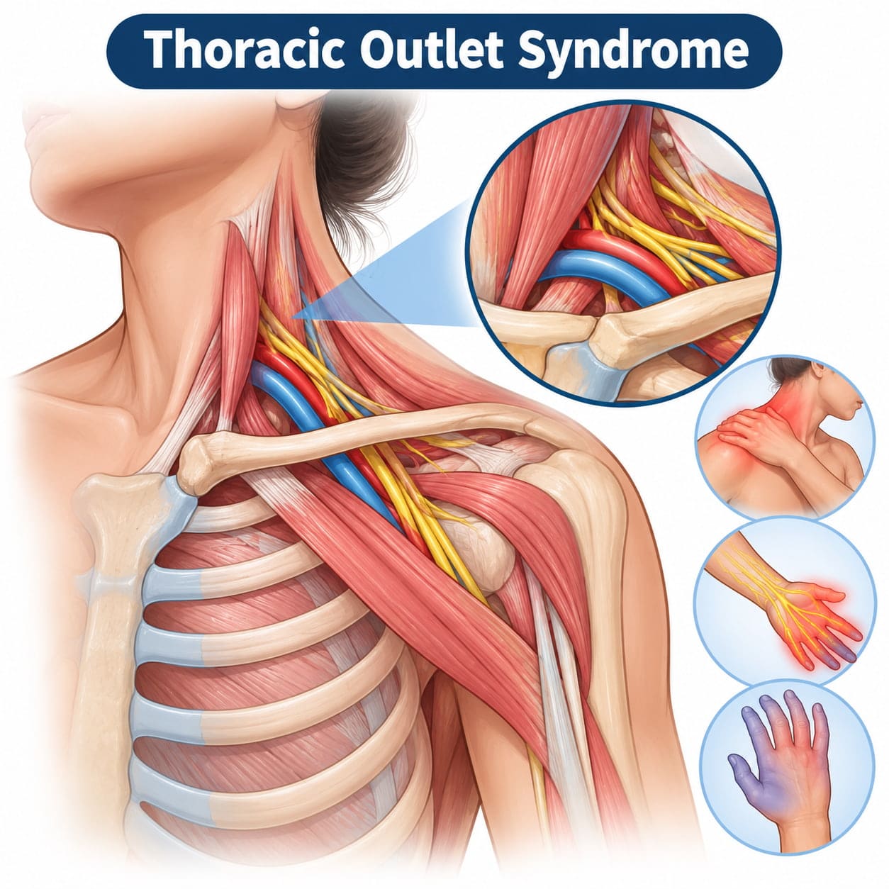
The thoracic outlet is the narrow space between the collarbone, or clavicle, and first rib where the brachial plexus, subclavian artery and subclavian vein pass from the neck into the upper extremity.
Thoracic outlet syndrome refers to a group of conditions in which these neurovascular structures become compressed within that space, producing neurological or vascular symptoms in the arm and hand.
The three main subtypes are neurogenic thoracic outlet syndrome, venous thoracic outlet syndrome and arterial thoracic outlet syndrome. Understanding which structures are compressed matters because each subtype has different risk factors, symptoms, and potential complications.
Common risk factors for thoracic outlet syndrome include anatomical variations and repetitive movements, but posture habits, previous injuries, occupation, sport and systemic factors can also contribute.
Recognising these risk factors can help patients, GPs and therapists consider TOS earlier when compatible symptoms appear and determine when specialist referral is appropriate.
Thoracic Outlet Syndrome Types and Their Risk Profiles
Neurogenic TOS is the most common form. Contemporary reviews describe neurogenic TOS as approximately 94–95% of cases, venous TOS as 3–4%, and arterial TOS as 1–2%.
The risk factors behind each subtype differ.
Neurogenic TOS involves compression of the brachial plexus and is more closely linked to posture, muscle imbalance, muscle tension, repetitive movement and congenital anatomical variations such as fibrous bands.
Venous TOS results from compression of the subclavian vein. It is often associated with strenuous overhead activity, repetitive movements and heavy use of the affected upper extremity.
Arterial TOS occurs because of compression of the subclavian artery and is more strongly associated with fixed bony abnormalities such as cervical ribs or anomalous first ribs.
Each subtype can also produce a different symptom pattern. Neurogenic TOS may cause numbness or tingling in the arm, arm pain and a weak grip. Venous thoracic outlet syndrome can cause arm swelling, heaviness and bluish discolouration of the hand, while arterial TOS may cause cold fingers, pallor or symptoms related to reduced blood flow.
These thoracic outlet symptoms can overlap, and some patients have more than one contributing risk factor. Careful subtype classification is therefore important before treatment is planned.
"Dr. Marco Scarci is a deeply knowledgeable, open-minded, and empathetic surgeon. He listens to details and solves problems to truly deliver the most optimal results. As a medical professional myself, I can tell you that finding a specialist like Dr. Scarci is very rare, and it is a privilege to have him in my corner."
Demographic Risk Factors: Who Is Most Commonly Affected?
Thoracic outlet syndrome can affect people of different ages, but some demographic patterns appear repeatedly across clinical series.
TOS is commonly diagnosed in younger and middle-aged adults, particularly between approximately 20 and 50 years of age, with many clinical series showing a concentration of cases in younger adults.
Women are diagnosed with neurogenic thoracic outlet syndrome more often than men. A UK multicentre surgical review involving 133 patients found that 65% were female.
The reasons for this difference are not fully established. Proposed contributors include differences in shoulder girdle anatomy, connective-tissue laxity, posture and other anatomical or physiological factors.
Venous TOS and Paget–von Schroetter syndrome, by contrast, are often seen in young, physically active people who perform repetitive overhead movements. Young male athletes and manual workers are commonly represented in venous TOS series.
Age and sex alone do not establish a diagnosis, but being in a higher-risk demographic can increase suspicion when compatible neurological or vascular symptoms appear.
Have a question about your diagnosis?
A consultation gives you a personalised plan — not a generic estimate.
Anatomical and Structural Risk Factors
Anything that narrows the thoracic outlet or changes the relationship between its structures can increase the likelihood of nerve compression, artery compression or vein stenosis.
Important areas where compression can occur include the scalene triangle, between the anterior and middle scalene muscles and the first rib; the costoclavicular space, between the clavicle and first rib; and the space beneath the pectoralis minor muscle.
Congenital anatomical risk factors include a cervical rib, anomalous first rib, fibrous bands arising from the cervical spine, unusual scalene muscle anatomy or hypertrophy, and congenital vascular abnormalities involving the subclavian vessels.
A large meta-analysis of 141 studies involving 77,924 participants found cervical ribs in 29.5% of symptomatic TOS patients compared with 1.1% of healthy individuals.
Cervical ribs are particularly important in vascular TOS. Fixed bony compression can damage the subclavian artery over time, potentially contributing to stenosis, aneurysm formation, thrombosis or distal embolisation.
Compression may also arise from abnormalities of the clavicle or first rib. Less commonly, masses or tumours in the lower neck or upper chest can narrow the thoracic outlet.
Acquired structural risk factors include malunited clavicle fractures, excessive fracture callus, scar tissue from previous surgery and deformity following trauma. Drooping of the shoulders associated with muscle wasting or substantial weight loss can also reduce the available space.
Targeted imaging with X-ray, CT, MRI or vascular imaging may help identify these structural problems after the clinical history and examination raise suspicion.

"After two years of being told my symptoms were anxiety, Mr Scarci identified the problem immediately. The surgery was straightforward and I was discharged after three days. I only wish I had found him sooner."
Posture, Muscle Imbalance and Lifestyle-Related Risk Factors
Poor posture is one of the most commonly discussed modifiable risk factors for neurogenic thoracic outlet syndrome.
Prolonged sitting, sustained forward head posture and rounded shoulders are common among people who spend long periods at desks, laptops or smartphones. Rounded shoulders, protracted scapulae and a flexed thoracic spine can reduce the available space in the costoclavicular and retropectoralis minor regions.
Over time, sustained compression can contribute to neurogenic symptoms, particularly when posture problems occur alongside anatomical susceptibility or repetitive arm use.
Muscle imbalance may involve tight pectoralis minor and scalene muscles, weak lower trapezius, serratus anterior or deep neck flexors, and excessive upper trapezius activity in people who frequently shrug or carry tension around the shoulder girdle.
Trigger points in the anterior and middle scalene muscles can also contribute to pain around the neck, shoulder and upper body.
Obesity may add downward loading around the shoulder girdle, while carrying heavy bags on one shoulder can further depress the clavicle and reduce the available space for nerves and blood vessels.
Many of these factors are modifiable. Targeted physical therapy, postural retraining, ergonomic changes and general conditioning form an important part of conservative treatment.
Want a specialist opinion on your condition?
Get a clear answer from a surgeon who will be with you throughout.
Occupational and Repetitive-Use Risk Factors
Many thoracic outlet problems are closely related to how the arms and shoulders are used repeatedly at work.
Occupations involving repetitive movements, sustained overhead arm positions, heavy lifting or prolonged posture can contribute to thoracic outlet compression over months or years.
Occupation | Primary Risk Mechanism |
Electricians and painters | Prolonged overhead arm movements |
Hairdressers | Arms held near shoulder height for extended periods |
Mechanics | Repetitive pulling and overhead reaching |
Warehouse operatives | Heavy lifting and carrying |
Healthcare workers | Patient handling and sustained positioning |
Desk work may create a different mechanism. Repetitive keyboard use combined with poor desk ergonomics, unsupported arms, forward head posture and shoulder protraction can gradually narrow the outlet and contribute to chronic pain or neurological symptoms.
Carrying heavy backpacks, tool belts, trays or shoulder bags can depress the clavicle and place additional tension on both the brachial plexus and subclavian vessels.
Heavy lifting and upper-body weight training can also reduce the available space when associated with muscle hypertrophy, repetitive strain or poor shoulder mechanics.
Early workplace assessment and ergonomic adjustment can reduce exposure to these factors. Occupational health input or physiotherapy may be particularly useful when symptoms are clearly linked to work posture or repetitive upper-extremity activity.
Sports and Activity-Related Risk Factors

Certain sports place repeated stress on the thoracic outlet because the arm is frequently elevated, abducted, externally rotated or placed under heavy load.
Sports commonly associated with TOS include swimming, baseball, tennis, volleyball, rowing, rock climbing and weightlifting.
A systematic review of Paget–von Schroetter syndrome in athletes identified 123 cases, with baseball accounting for 26.8% and weightlifting for 19%.
Paget–von Schroetter syndrome is effort thrombosis of the axillo-subclavian vein and represents a form of venous thoracic outlet syndrome. Repetitive overhead or pulling movements create chronic compression and microtrauma to the subclavian vein, eventually contributing to thrombosis in susceptible individuals.
Typical presentations include a swimmer, rower, climber, or weightlifter developing sudden unilateral arm swelling, heaviness, prominent veins, or bluish discolouration after strenuous activity.
These symptoms can indicate venous obstruction and should not be managed as a routine muscular injury. Acute venous thrombosis requires urgent vascular assessment because complications can include pulmonary embolism.
Not sure about your treatment options?
Mr Scarci provides expert consultations typically within one week of contact.
Trauma, Previous Injury and Post-Surgical Changes
Previous injury to the neck, shoulder girdle or upper chest can alter local anatomy and predispose to thoracic outlet syndrome later.
TOS sometimes develops when an underlying anatomical predisposition is made symptomatic by trauma.
Relevant injuries include whiplash from road traffic collisions, falls directly onto the shoulder, high-energy sports injuries, clavicle fractures and first rib fractures.
Sudden trauma can create swelling, scarring or structural changes around the thoracic outlet. A clavicle fracture that heals with malunion or excessive callus can narrow the costoclavicular space. Plate-and-screw fixation near the outlet may also alter local anatomy in selected cases.
Repeated injuries to the same area can compound these effects.
Post-surgical scarring or postural changes following lung, breast, cardiac or neck surgery can also alter the outlet. New neurological or vascular symptoms affecting an upper extremity after surgery should therefore be assessed carefully rather than automatically attributed to routine postoperative discomfort.
A thorough medical history should include previous fractures, operations and trauma involving the neck, clavicle, shoulder and upper chest because these structural contributors may be overlooked.
Systemic and Physiological Risk Factors
Systemic factors do not always directly cause thoracic outlet compression, but they can increase susceptibility or make the consequences of compression more significant.
Pregnancy may temporarily alter posture, fluid balance and joint laxity. Weight gain and changes in the shoulder girdle can exacerbate an already narrow outlet and increase neurogenic symptoms.
Obesity and generalised ligamentous laxity or hypermobility can similarly alter shoulder positioning and thoracic outlet mechanics.
For venous TOS and Paget–von Schroetter syndrome, additional thrombotic risk factors can compound the effects of mechanical compression.
These can include inherited thrombophilia such as factor V Leiden or protein C or S deficiency, oral contraceptive use, smoking and prolonged immobility.
These factors do not necessarily cause TOS by themselves. Instead, they can lower the threshold at which anatomical or activity-related compression becomes clinically important and increase the likelihood of blood clots or long-term vascular complications.
How Risk Factors Are Identified in Practice
Risk factors for thoracic outlet syndrome are often uncovered through careful history-taking and physical examination before advanced imaging is requested.
A detailed medical history explores occupation, sports participation, previous injuries, operations, systemic illness and symptom triggers related to arm position or load.
The clinician may ask whether symptoms worsen when the arms are raised, when carrying bags, during exercise or after prolonged desk work. Posture, sleep position and repetitive tasks may also be relevant.
Conditions that produce similar symptoms should be considered during this process. Cervical radiculopathy, carpal tunnel syndrome, rotator cuff disease, peripheral nerve entrapment and other musculoskeletal or neurological conditions can resemble TOS. Careful assessment of conditions that mimic TOS is therefore an important part of avoiding misdiagnosis.
Physical examination may include inspection of posture and shoulder height, assessment for rounded shoulders, palpation of the supraclavicular region, evaluation of hand muscle bulk, neurological examination and comparison of pulses or blood pressure between arms.
Provocation tests may also be used to reproduce symptoms or assess positional vascular changes. These can include Adson’s test, the Roos or elevated arm stress test, Wright’s test and the costoclavicular manoeuvre.
Do not interpret these tests in isolation, as individual provocative manoeuvres have limited diagnostic accuracy. Consider their findings alongside symptoms, examination, imaging, and the patient’s overall risk profile.
Imaging Correlates of Risk Factors
Imaging helps confirm anatomical abnormalities and identify vascular consequences of thoracic outlet compression.
Imaging Modality | Primary Use in TOS |
Plain X-ray | Detect cervical ribs, anomalous first ribs and old clavicle fractures |
Duplex ultrasound | Assess dynamic vessel compression and detect venous thrombosis |
CT angiography | Map the thoracic outlet and assess arterial anatomy or aneurysm |
MR angiography | Assess vascular anatomy without ionising radiation |
MRI | Evaluate soft-tissue structures and brachial plexus abnormalities |
Ultrasound and duplex scanning of the subclavian vein and subclavian artery can be performed with the arm in different positions to assess dynamic compression that may be less obvious on static imaging.
CT angiography and MR angiography become particularly relevant when arterial TOS, venous TOS or thoracic outlet decompression surgery is being considered.
MRI may be useful for evaluating soft tissues, brachial plexus pathology and alternative neurological or musculoskeletal diagnoses.
Importantly, anatomical abnormalities do not automatically mean a patient has symptomatic TOS. Cervical ribs, fibrous bands and other variants may remain asymptomatic throughout life.
Imaging therefore needs to be interpreted alongside the medical history, physical examination, symptoms and functional limitations rather than used as a standalone diagnosis.
Have a question about your diagnosis?
A consultation gives you a personalised plan — not a generic estimate.
Risk Factors for Chronic Pain and Long-Term Complications
Not every case of thoracic outlet compression leads to chronic pain or disability, but delayed diagnosis and ongoing exposure to provoking activities can increase the likelihood of long-term problems.
In neurogenic TOS, prolonged brachial plexus compression can contribute to progressive hand muscle wasting, permanent sensory change, weak grip, impaired hand function and chronic pain.
Long-standing neurological symptoms can become more difficult to treat when significant nerve dysfunction is already established.
Arterial TOS carries different risks. Persistent compression of the subclavian artery can cause arterial damage, stenosis, aneurysm formation or distal embolisation. In severe cases, reduced blood flow can progress to critical limb ischaemia and require complex vascular reconstruction.
Venous TOS and recurrent Paget–von Schroetter syndrome can cause chronic venous obstruction, persistent swelling, collateral vein formation, and functional limitation of the affected arm.
Cold fingers, marked pallor, bluish hand discolouration or significant unilateral swelling should not be dismissed as minor symptoms because they may indicate vascular compromise.
Earlier assessment allows the underlying mechanical and vascular factors to be addressed before irreversible nerve or vessel damage develops.
Modifiable vs Non-Modifiable Risk Factors
Some thoracic outlet syndrome risk factors cannot be changed, while others can be reduced through rehabilitation, lifestyle changes or altered activity.
Non-modifiable factors include congenital cervical ribs or other fixed anatomical abnormalities, permanent structural changes after major trauma, and certain demographic or connective-tissue characteristics.
Modifiable factors include poor posture, forward head posture, repetitive overhead work, asymmetric bag carrying, smoking, excess weight, poor conditioning of shoulder muscles and repeated activity that overloads the thoracic outlet.
Addressing modifiable factors is an important part of treatment even when a fixed anatomical abnormality is also present.
A prospective observational cohort of patients with neurogenic TOS found that 40 patients improved with physical therapy alone, showing that structured rehabilitation can be effective for a meaningful proportion of appropriately selected patients.
Reducing modifiable risks can also improve surgical preparation and long-term symptom control when surgery eventually becomes necessary.
Conservative Management Focused on Risk Reduction
Conservative treatment is generally the first step for neurogenic TOS unless there is progressive neurological loss or another reason for earlier intervention.
Physiotherapy programmes commonly focus on correcting rounded shoulders and forward head posture, improving scapular control, strengthening the lower trapezius, serratus anterior and other scapular stabilisers, and stretching tight pectoralis minor and scalene muscles.
Breathing mechanics can also be addressed because overuse of the accessory breathing muscles may increase scalene tension and first-rib elevation.
Treatment may additionally target trigger points and muscle tension using appropriate manual therapeutic techniques.
Ergonomic assessment of workstations and daily activities can reduce sustained arm elevation, shoulder loading and repetitive movements. Pain-management strategies, graded activity and modification of provoking tasks aim to reduce symptoms while function is rebuilt.
Lifestyle recommendations may include weight management, smoking cessation, regular breaks from repetitive activity and a graded return to sport using improved technique.
Conservative treatment should still be monitored. Persistent weakness, progressive neurological deficit, arm swelling or vascular changes require reassessment rather than indefinite physiotherapy.
When Risk Factors Indicate Specialist or Surgical Evaluation
Certain combinations of risk factors and symptoms should prompt earlier referral to a specialist rather than prolonged conservative care alone.
A known cervical rib combined with a cold or pale hand, reduced pulse or evidence of arterial insufficiency raises concern for arterial TOS. A young overhead athlete with sudden unilateral arm swelling may have Paget–von Schroetter syndrome and requires urgent investigation for venous thrombosis.
Progressive neurological deficit in a patient with clavicle malunion, recurrent venous thrombosis despite appropriate management, or persistent functional impairment despite structured physiotherapy can also justify specialist evaluation.
Acute venous thrombosis may require thrombolysis or anticoagulation before definitive decompression is considered.
Surgical options can include first rib resection, scalenectomy, cervical rib removal and thoracic outlet decompression, depending on which structures are causing compression.
A UK multicentre review involving 133 surgically treated patients reported 89% symptom resolution, although outcomes varied by subtype and neurogenic TOS remains particularly dependent on careful patient selection.
In Mr Marco Scarci’s practice, the clinical history, anatomical findings, imaging and response to conservative treatment are considered together before surgery is recommended. As a private thoracic surgeon, he assesses and manages thoracic outlet syndrome and other complex chest conditions.
Want a specialist opinion on your condition?
Get a clear answer from a surgeon who will be with you throughout.
Frequently Asked Questions
Can I develop thoracic outlet syndrome without playing sport or doing physical work?
Yes. Thoracic outlet syndrome (TOS), particularly neurogenic TOS, can also affect people with sedentary lifestyles. Prolonged desk work and sustained head-and-shoulder positions may contribute to symptoms in some people. Factors such as pregnancy, hypermobility or anatomical variations can also play a role.
Which symptoms need urgent medical assessment?
Seek urgent assessment for sudden arm swelling, blue or very pale skin, a clear temperature difference between arms, or severe pain or weakness. These symptoms can indicate a vascular problem and should not be managed at home without medical advice.
Does a cervical rib mean I will develop TOS?
No. Many people with a cervical rib never develop symptoms. However, a cervical rib may increase the likelihood of TOS when combined with factors such as trauma, repetitive overhead activity or vascular abnormalities. Speak with a clinician if you have compatible symptoms.
Can treatment reduce symptoms without surgery?
Often, neurogenic TOS associated with posture or muscle imbalance is initially managed with targeted physiotherapy and activity adjustments. Surgery may be considered where symptoms persist or where there is a significant structural or vascular cause. The right approach depends on the individual assessment.
Should relatives be screened for TOS?
Routine screening is not usually needed. TOS is not generally considered strongly hereditary. A clinician may consider assessment for relatives who have relevant symptoms and a known family history of anatomical or vascular variations.
