

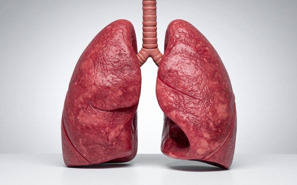
Distinguishing between lung cancer and mesothelioma affects diagnosis accuracy, treatment planning, and patient outcomes. While both conditions can affect the chest area and share common symptoms such as persistent cough, chest pain, and fatigue, they develop in different locations and require distinct surgical approaches. Understanding these differences is crucial for patients with respiratory symptoms or an asbestos exposure history.
This guide compares lung cancer and mesothelioma in terms of causes, symptoms, diagnosis, treatment options, and prognosis. It is intended for patients, families, and healthcare professionals seeking to understand the key differences and implications for care. By clarifying how these diseases differ, this resource aims to support informed decision-making and improve outcomes for those affected.
Lung cancer forms inside the lung tissue as defined tumours, while mesothelioma spreads across the lining of the lung.
Smoking is the leading cause of lung cancer, whereas mesothelioma is mainly linked to asbestos exposure, often decades before symptoms appear.
Diagnosis and treatment differ because lung cancer can often be surgically targeted as a clear tumour, while mesothelioma usually requires specialist biopsy, complex surgery, and multimodal care.
Anyone with persistent cough, chest pain, breathlessness, or a history of asbestos exposure should seek specialist assessment, as earlier diagnosis can improve treatment options and outcomes.
Lung Cancer vs Mesothelioma: Key Medical Differences
The fundamental distinction between mesothelioma and lung cancer lies in where the cancer develops and how it grows.
Mesothelioma is a rare cancer almost exclusively caused by asbestos exposure, developing in the lining of the lungs (pleura) or other organs. Malignant pleural mesothelioma develops from the mesothelial cells lining the lungs, spreading as a diffuse rind across the pleural surface rather than forming a single solid mass.
Lung cancer is a malignancy that develops in the tissues of the lung, most commonly caused by smoking, radon, and asbestos. Lung cancer forms inside lung tissue, within the bronchi, bronchioles, or alveoli, creating discrete tumour masses that can often be surgically targeted with defined margins.
The difference in incidence is also striking. In the UK, lung cancer affects over 43,000 patients per year, compared with roughly 2,500 cases of malignant mesothelioma.
Both mesothelioma and lung cancer require specialised thoracic surgical expertise, but the operative approaches differ significantly. Lung cancer surgery typically aims to remove a defined mass with clear margins, while mesothelioma surgery must address widespread disease across the pleural surface, making complete resection far more challenging.
Not sure about your treatment options?
Mr Scarci provides expert consultations typically within one week of contact.
Location and Development Patterns
How Lung Cancer Develops
Lung cancer forms inside the lung parenchyma. Cancer cells arise within the bronchial tree or peripheral lung tissue and grow as defined masses that compress surrounding structures. The most common types of non-small cell lung cancer include adenocarcinoma, squamous cell carcinoma, and large cell carcinoma, which together account for approximately 80–85% of lung cancer cases. Small cell lung cancer makes up the remaining 10–15%.
Because lung cancer forms discrete tumours, CT imaging can identify their location precisely, and surgical resection can be planned with clear anatomical targets. Tumours may originate centrally in major bronchi or peripherally in the lung parenchyma. As they grow, lung cancer cells may invade the chest wall, spread to lymph nodes along predictable anatomical pathways, or develop into metastatic cancer affecting distant organs.
Lung cancer can take 15–30 years to develop, which is why screening programmes focus on high-risk populations to catch disease at earlier, more treatable stages and address how long lung cancer can remain undetected.
How Mesothelioma Develops
Mesothelioma develops in an entirely different pattern. Rather than forming a solid mass within lung tissue, pleural mesothelioma starts in the mesothelial cells lining the chest cavity. It spreads diffusely across both the visceral and parietal pleura, creating a thick rind of tumor tissue that encases the lung.
This diffuse growth pattern means mesothelioma forms along surfaces rather than invading as a discrete mass. It can infiltrate the diaphragm, chest wall, and pericardium, which is why pericardial mesothelioma and peritoneal mesothelioma also occur, though pleural disease is by far the most common.
Mesothelioma typically develops 10–50 years after asbestos exposure, with the latency period most commonly falling between 20 and 50 years. This extended timeline between cause and diagnosis means many patients are elderly at presentation; over 63% of UK cases are diagnosed in people aged 75 or older.
The diffuse nature of mesothelioma cells makes achieving negative surgical margins extremely difficult, fundamentally changing the surgical approach compared with lung cancer.
"After two years of being told my symptoms were anxiety, Mr Scarci identified the problem immediately. The surgery was straightforward and I was discharged after three days. I only wish I had found him sooner."
Causes and Risk Factors
Lung Cancer Risk Factors
The primary cause of lung cancer is smoking, which accounts for an estimated 85–90% of lung cancer cases. However, lung cancer can develop from multiple causes, including radon gas exposure, air pollution, occupational hazards, and family history. Asbestos lung cancer is also a recognised risk factor, asbestos is one of several environmental causes that can trigger malignant changes in lung tissue.
The combined effect of risk factors can be dramatic: a person exposed to asbestos and smoking is 50–90 times more likely to develop lung cancer than someone with neither exposure. This synergistic relationship makes smoking cessation particularly urgent for anyone with occupational asbestos exposure.
Other occupational exposures across various industries, inherited gene mutations (such as EGFR and ALK), and previous lung disease all contribute to overall cancer risk. Each type of lung cancer has slightly different risk factor profiles, squamous cell carcinoma and small cell lung cancer are most strongly linked to smoking, while adenocarcinoma is the most common subtype in non-smokers.
Mesothelioma Risk Factors
The risk factor profile for mesothelioma is far more specific. Asbestos exposure causes nearly all mesothelioma cases. Unlike lung cancer, where smoking is the dominant cause, and asbestos is one of many contributors, mesothelioma is almost exclusively an asbestos-related disease.
Asbestos exposure is commonly associated with occupations such as construction, shipbuilding, insulation installation, and industrial manufacturing. When disturbed, asbestos fibres become airborne and can be inhaled, embedding in the pleural lining where they cause chronic inflammation and eventually malignant transformation over decades.
The latency period for mesothelioma is typically 20–50 years after asbestos exposure, explaining why incidence peaks in the 80–84 age group despite the substance being heavily regulated since the 1990s.
Genetic mutations in the BAP1 gene can increase susceptibility to mesothelioma. Carriers of germline BAP1 mutations show markedly different outcomes, with approximately 47% five-year survival compared with 6–7% in the general mesothelioma population. The International Mesothelioma Interest Group continues to study these genetic factors to identify surveillance strategies for at-risk individuals.
Have a question about your diagnosis?
A consultation gives you a personalised plan — not a generic estimate.
Clinical Presentation and Symptoms
Lung Cancer Symptoms
Lung cancer symptoms vary depending on tumour location and size. The most common presentations include:
Persistent cough that lasts more than three weeks
Haemoptysis (coughing blood)
Chest pain and shortness of breath
Wheezing or stridor, particularly with central tumours
Systemic symptoms including weight loss, fatigue, and loss of appetite
Symptoms often appear at advanced stages for both cancers, which is why early detection through lung cancer screening programmes is so important. Peripheral tumours may remain entirely asymptomatic until they have grown significantly or spread to lymph nodes.
Mesothelioma Symptoms
Mesothelioma commonly presents with pleural effusion, a buildup of fluid between the lung and chest wall that causes progressive breathlessness. The most frequent symptoms include:
Pleuritic chest pain (sharp, worsened by breathing)
Progressive dyspnoea from fluid buildup in the chest cavity
Dry cough and chest tightness
Late-stage symptoms including severe chest wall pain, weight loss, and fatigue
Both cancers share similar symptoms such as persistent cough, chest pain, and fatigue, which is one reason clinicians can initially misdiagnose mesothelioma as a benign pleural condition or even as lung cancer. Shortness of breath is a common symptom in both cancers, and both cancers can lead to pleural effusion, making clinical differentiation challenging without tissue sampling.
Because pleural mesothelioma affects the lining rather than the lung itself, early symptoms can be subtle and easily attributed to other conditions, contributing to the characteristically late-stage presentation.
"After multiple failed consultations elsewhere, Mr Scarci identified exactly what was wrong and operated within two weeks. Recovery was smooth and I'm back to normal life."
Diagnostic Approaches
Lung Cancer Diagnosis
Diagnosing lung cancer typically follows a structured pathway. Diagnostic techniques for both cancers include medical history, imaging, and biopsies, but the specific tools differ.
First-line imaging: Contrast-enhanced CT chest scans are standard for evaluating suspected lung cancer
Tissue sampling: Bronchoscopy allows direct visualisation and biopsy of central lesions, while CT-guided percutaneous biopsy targets peripheral nodules
Staging: PET-CT scans assess the extent of disease spread and guide the treatment plan
Molecular testing: EGFR, ALK, ROS1, KRAS, and PD-L1 testing is now front-loaded to identify targetable driver mutations
Imaging tests include X-rays, CT or PET scans for diagnosis. NHS lung cancer screening using low-dose CT now detects approximately 75.7% of cancers at Stage I or II, compared with fewer than 30% at early stages without screening, a transformative shift in how doctors diagnose and treat the disease.
Mesothelioma Diagnosis
Diagnosing mesothelioma is considerably more challenging. Pleural fluid cytology, the analysis of fluid drained from the chest, has limited sensitivity and is often non-diagnostic.
Gold standard tissue diagnosis: Thoracoscopy (VATS) allows direct visualisation and biopsy of the pleural surface, providing adequate tumor tissue for analysis
Immunohistochemistry: Markers including calretinin, WT-1, CK5/6, D2-40, and loss of BAP1 nuclear staining are essential for definitive diagnosis and distinguishing mesothelioma from other malignancies
Subtype classification: Histological subtyping (epithelioid, biphasic, or sarcomatoid) directly impacts prognosis and treatment decisions
Specialist pathology review: Given diagnostic complexity, expert pathological assessment is critical for accurate mesothelioma diagnosis
CT scans are standard for evaluating malignant pleural mesothelioma, sometimes supplemented with MRI or other cross-sectional imaging or PET imaging. A biopsy is essential for confirming a mesothelioma diagnosis, relying on imaging alone risks significant diagnostic error. The difficulty in distinguishing mesothelioma from other pleural diseases or even from advanced lung cancer underscores the importance of referral to a mesothelioma specialist with access to expert pathology services.
Want a specialist opinion on your condition?
Get a clear answer from a surgeon who will be with you throughout.
Surgical Treatment Options
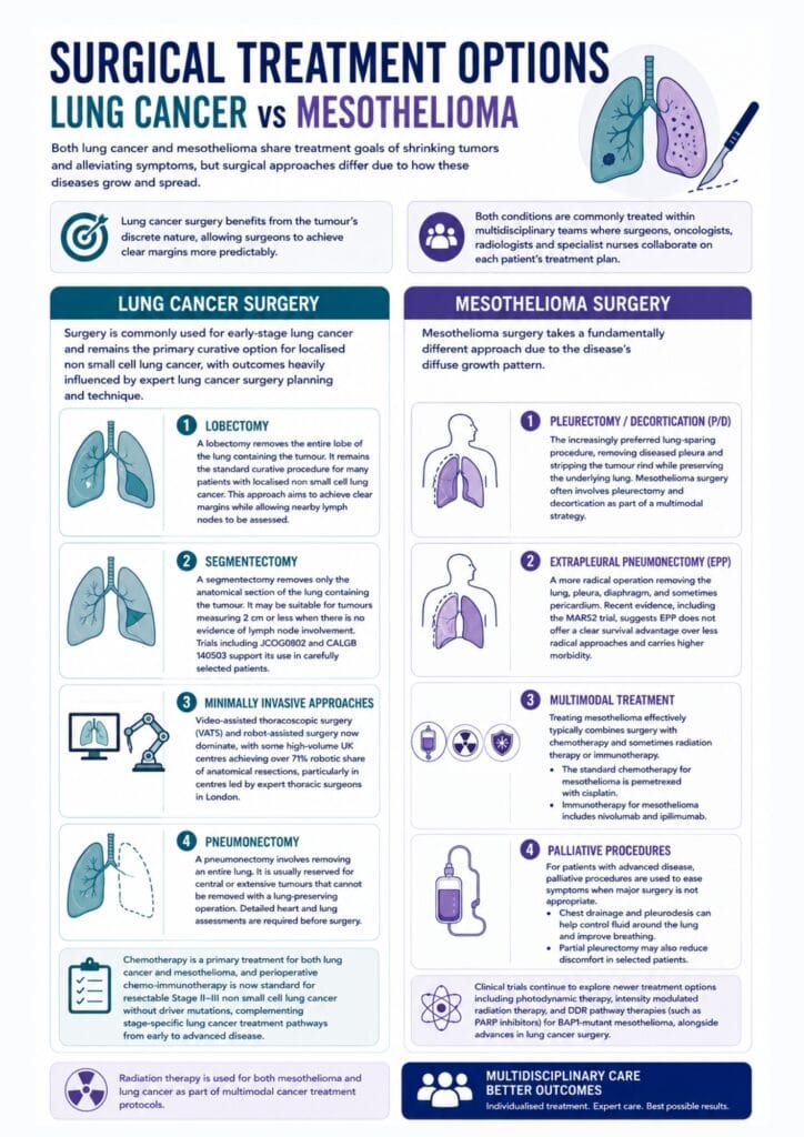
Lung Cancer Surgery
Surgery is commonly used for early-stage lung cancer and remains the primary curative option for localised non-small-cell lung cancer, with outcomes heavily influenced by expert lung cancer surgery planning and technique.
Lobectomy
A lobectomy removes the entire lobe of the lung containing the tumour. It remains the standard curative procedure for many patients with localised non-small-cell lung cancer. This approach aims to achieve clear margins while allowing nearby lymph nodes to be assessed.
Segmentectomy
A segmentectomy removes only the anatomical section of the lung containing the tumour. It may be suitable for tumours measuring 2 cm or less when there is no evidence of lymph node involvement. Trials including JCOG0802 and CALGB 140503 support its use in carefully selected patients.
Minimally invasive approaches
Video-assisted thoracoscopic surgery and robot-assisted surgery now dominate, with some high-volume UK centres achieving over 71% robotic share of anatomical resections, particularly in centres led by expert thoracic surgeons in London.
Pneumonectomy
A pneumonectomy involves removing an entire lung. It is usually reserved for central or extensive tumours that cannot be removed with a lung-preserving operation. Detailed heart and lung assessments are required before surgery. Reserved for central or extensive tumours where lung-preserving options are not feasible
Both lung cancer and mesothelioma share treatment goals of shrinking tumours and alleviating symptoms, but lung cancer surgery benefits from the tumour’s discrete nature, allowing surgeons to achieve clear margins more predictably.
Chemotherapy is a primary treatment for both lung cancer and mesothelioma, and perioperative chemo-immunotherapy is now standard for resectable Stage II–III non-small cell lung cancer without driver mutations, complementing stage-specific lung cancer treatment pathways from early to advanced disease. Radiation therapy is used for both mesothelioma and lung cancer as part of multimodal cancer treatment protocols.
Mesothelioma Surgery
Mesothelioma surgery takes a fundamentally different approach due to the disease’s diffuse growth pattern.
Pleurectomy/decortication (P/D)
The increasingly preferred lung-sparing procedure, removing diseased pleura and stripping the tumour rind while preserving the underlying lung. Mesothelioma surgery often involves pleurectomy and decortication as part of a multimodal strategy.
Extrapleural pneumonectomy (EPP)
A more radical operation removing the lung, pleura, diaphragm, and sometimes pericardium. Recent evidence, including the MARS2 trial, suggests EPP does not offer a clear survival advantage over less radical approaches and carries higher morbidity.
Multimodal treatment
Treating mesothelioma effectively typically combines surgery with chemotherapy and sometimes radiation therapy or immunotherapy. The standard chemotherapy for mesothelioma is pemetrexed with cisplatin. Immunotherapy for mesothelioma includes nivolumab and ipilimumab.
Palliative procedures
For patients with advanced disease, palliative procedures are used to ease symptoms when major surgery is not appropriate. Chest drainage and pleurodesis can help control fluid around the lung and improve breathing. Partial pleurectomy may also reduce discomfort in selected patients.
Clinical trials continue to explore newer treatment options, including photodynamic therapy, intensity-modulated radiation therapy, and DDR pathway therapies (such as PARP inhibitors) for BAP1-mutant mesothelioma, alongside advances in lung cancer surgery. Both conditions are commonly treated within multidisciplinary team settings where surgeons, oncologists, and specialist nurses collaborate on each patient’s treatment plan.
Prognosis and Outcomes
The prognostic outlook for these two conditions differs substantially.
Lung Cancer Prognosis
The 5-year survival rate for lung cancer is 28%. About 55% of patients survive at least one year. Lung cancer prognosis varies widely depending on type and stage. Early-stage disease treated surgically offers excellent outcomes, with five-year survival exceeding 90% for small peripheral tumours operated on in high-volume specialist centres. However, lung cancer diagnosed at an advanced stage carries a poor prognosis, with Stage IV median survival under one year. This is an aggressive cancer when detected late, but both lung cancer and mesothelioma can have better outcomes with early diagnosis.
Mesothelioma Prognosis
The 5-year survival rate for mesothelioma is 12%. About 53% of mesothelioma patients survive at least one year. Median survival for mesothelioma is approximately one year.
Mesothelioma prognosis depends heavily on histological subtype. Epithelioid malignant pleural mesothelioma, the most common subtype, carries a median survival of 14–19 months and responds best to multimodal treatment. Sarcomatoid and biphasic subtypes have worse outcomes, with sarcomatoid disease often showing median survival under 12 months and a particularly poor prognosis.
Life expectancy for both conditions is influenced by:
Stage at diagnosis
Histological cell type
Patient fitness and performance status
Access to specialist care at high-volume centres
Molecular and genetic markers
For peritoneal mesothelioma treated with aggressive cytoreductive surgery plus HIPEC in selected centres, five-year survival can reach approximately 65%, dramatically better than pleural disease, though this applies only to carefully selected patients, similar to how selected patients with pneumothorax benefit from tailored pneumothorax surgery procedures and expectations.
Mesothelioma is rare, with around 2,500-3,000 deaths per year in the United States, yet its impact on affected individuals and families is profound given its typically aggressive course and limited treatment options.
Not sure about your treatment options?
Mr Scarci provides expert consultations typically within one week of contact.
Legal and Financial Considerations
Patients diagnosed with asbestos-related lung cancer or mesothelioma may be entitled to compensation to help cover medical bills, lost wages, travel expenses, and other costs linked to their illness.
Legal action may be taken against employers, manufacturers, property owners, or other companies responsible for asbestos exposure. Access to financial support can reduce some of the pressure on patients and families while also making it easier to pursue specialist care, second opinions, and appropriate cancer treatment.
When to Seek Specialist Consultation

Any patient with persistent respiratory symptoms, a persistent cough lasting more than three weeks, unexplained breathlessness, chest pain, or haemoptysis should be evaluated promptly. Early referral to a specialist can make a meaningful difference to outcomes.
Patients with a history of asbestos exposure should always mention this to their doctors, even if exposure occurred decades ago. Given the 20–50 year latency period, even subtle pleural thickening or unexplained pleural effusion in someone with occupational exposure warrants specialist review.
For complex cases, particularly where there is diagnostic uncertainty between lung cancer and mesothelioma or where advanced surgical techniques may be required, seeking a second opinion from a specialist thoracic surgeon is strongly recommended. High-volume centres with multidisciplinary teams consistently achieve better outcomes for both conditions.
Virtual consultations can provide an initial assessment for patients who cannot easily travel, including international patients, and some choose to access private thoracic surgery in London for faster treatment. However, definitive diagnostic procedures and surgery should be performed at centres with thoracic surgery accreditation, experienced pathology services, and robust postoperative support, particularly for mesothelioma, where surgical complexity and the need for multimodal treatment demand specialist expertise.
