

Thymoma is a rare tumour arising from the thymus gland behind the breastbone. Because it grows slowly and sits deep inside the chest, early thymoma symptoms are often subtle, vague, or completely absent.
This guide explains the warning signs worth knowing, from persistent chest symptoms to immune-related conditions like myasthenia gravis, and outlines when to seek specialist assessment.
Many people with thymoma experience no initial symptoms. The tumour is frequently discovered incidentally on a chest X-ray or CT scan performed for an unrelated reason.
The most common early warning signs include a persistent dry cough, vague chest pressure or pain, and mild shortness of breath on exertion.
Thymoma is a rare cancer of the thymus gland in the anterior mediastinum and is frequently associated with autoimmune diseases, particularly myasthenia gravis, which affects up to 50% of thymoma patients.
New, otherwise unexplained muscle weakness, especially drooping eyelids, double vision, or difficulty chewing and swallowing, should prompt urgent medical review.
Understanding the Thymus and Thymoma
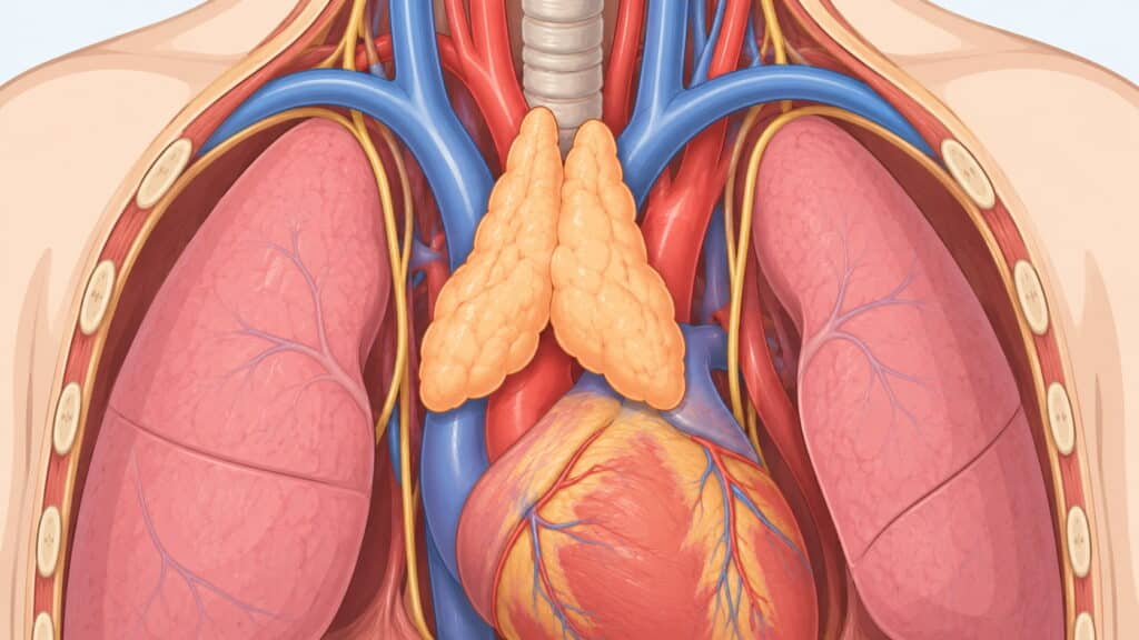
The thymus is a small gland situated behind the sternum, between the lungs, in an area called the anterior mediastinum. It is most active during childhood and adolescence, when it helps produce white blood cells known as T-lymphocytes, cells that play a central role in helping the immune system fight infection and disease.
Thymoma is a tumour that develops from the thymic epithelial cells, the lining cells of the gland. It is considered a rare cancer, with an incidence in the UK and Ireland of roughly 1.1 cases per million people per year.
Thymomas are classified into two main types: A and B. The WHO classifies thymomas into six histological types overall.
Type A thymomas usually grow slowly and rarely spread beyond the thymus, while Type B thymomas, including the predominantly cortical thymoma subtype, can grow more quickly and may spread to the lungs or nearby fatty tissue.
It is important to distinguish between thymoma and thymic carcinoma. Thymic carcinoma is rarer, tends to grow more aggressively and is more likely to metastasise to bones and liver. In contrast, thymomas generally remain confined to the chest for longer periods.
Together, thymoma and thymic carcinoma represent the main thymic epithelial tumours. Understanding the thymus also helps explain why these thymic tumours are so closely linked with autoimmune diseases and other paraneoplastic syndromes that disrupt the body’s ability to distinguish between healthy cells and tumour cells.
Not sure about your treatment options?
Mr Scarci provides expert consultations typically within one week of contact.
How and When Thymoma Is Usually Detected
A striking feature of thymoma is how often it is found by accident. Research involving large patient cohorts shows that roughly 43% of thymoma patients are completely asymptomatic at diagnosis.
Thymomas are often discovered incidentally during imaging tests. This might include a routine chest X-ray before planned surgery or a CT scan ordered for a persistent chest infection or suspected lung disease.
The increasing use of CT scans in modern medicine has led to more incidental findings, identifying small thymic malignancies before they cause noticeable problems. However, the absence of symptoms does not mean the tumour is harmless.
Even an apparently quiet early-stage thymoma requires specialist assessment because cancer cells can invade surrounding tissues or trigger immune complications without producing obvious chest symptoms.
Early referral to a thoracic surgeon allows careful evaluation and discussion of the safest, least invasive treatment options, ideally before the tumour has a chance to grow into nearby tissues or nearby organs.
"I was diagnosed in March with a rare condition. My local hospital said it was inoperable due to the size of the mass. Mr Scarci reassured me the thymoma was resectable. I was booked for surgery the following week where he successfully removed the entire tumour."
Early Local Symptoms in the Chest
When thymoma symptoms appear, they usually result from mass effect, with the tumour pressing on nearby chest structures in the mediastinum. The health effects of thymoma can therefore include respiratory compromise and functional decline.
The most common early local signs include:
Persistent dry or irritating cough that does not settle with usual treatments
Vague chest discomfort, chest pressure or dull chest pain behind the breastbone
Shortness of breath or tightness, especially on exertion or when lying flat
A feeling of fullness in the upper chest or difficulty taking a deep breath
Less common but still relevant early signs include hoarseness of the voice, which can develop from pressure on the recurrent laryngeal nerve, and mild difficulty swallowing, known as dysphagia, from pressure on the oesophagus. Symptoms may include hoarseness and a persistent cough that patients dismiss for months.
These early symptoms are non-specific. Thymoma symptoms may overlap with lung cancer, autoimmune diseases and other chest conditions, and they are frequently mistaken for asthma, chest infection, heartburn or musculoskeletal pain.
Any chest pain associated with breathlessness, unexplained weight loss or new fatigue warrants prompt GP review and, if needed, referral for chest imaging.
Have a question about your diagnosis?
A consultation gives you a personalised plan — not a generic estimate.
Symptoms Related to Pressure on the Superior Vena Cava
The superior vena cava is a large vein that carries blood from the head, neck, upper body, upper chest and arms back to the heart. It runs very close to the thymus, and these blood vessels can be compressed by a growing tumour.
When a thymoma compresses this vein, it can cause superior vena cava syndrome, which can occasionally be an early presentation in larger or more invasive thymic cancers.
Characteristic symptoms include:
Swelling or oedema of the face, neck and upper chest
Puffiness around the eyes, especially in the morning
Prominent, enlarged visible veins on the chest wall or neck
Shortness of breath, cough or a feeling of head pressure when bending forward or lying flat
Headache, dizziness or a sensation of fullness in the head
Superior vena cava syndrome can be serious and occasionally life-threatening. If these symptoms appear suddenly or worsen quickly, urgent assessment in an emergency department is required.
Rapid imaging with a CT scan and input from a thoracic oncology and thoracic surgery team may be needed to stabilise the patient and plan definitive cancer treatment.
"Dr. Marco Scarci is a deeply knowledgeable, open-minded, and empathetic surgeon. He listens to details and solves problems to truly deliver the most optimal results. As a medical professional myself, I can tell you that finding a specialist like Dr. Scarci is very rare, and it is a privilege to have him in my corner."
Early Immune-Related Symptoms: Myasthenia Gravis
Thymoma is strongly associated with autoimmune diseases, where the immune system mistakenly attacks healthy tissue instead of protecting it. The most common is myasthenia gravis, a condition that affects communication between nerves and muscles at the neuromuscular junction.
Myasthenia gravis is the most common autoimmune disease linked to thymomas. Approximately 30% to 50% of thymoma patients develop myasthenia gravis, and conversely, about 10% to 15% of people diagnosed with myasthenia gravis are found to have a thymoma.
In some patients, symptoms of myasthenia gravis appear months or even years before the thymic tumour is identified on imaging.
Early warning signs include:
Drooping eyelids (ptosis), often worse towards the end of the day
Double vision, especially after looking up or to the side for a prolonged period
Fluctuating muscle weakness that improves with rest and worsens with activity
Difficulty chewing tough foods, swallowing liquids or speaking clearly by evening
Weakness in the neck muscles causing the head to feel heavy
Myasthenia gravis causes fluctuating muscle weakness that worsens with activity because antibodies interfere with nerve-to-muscle communication. This makes it a classic paraneoplastic syndrome associated with thymoma.
The close relationship between thymoma and myasthenia gravis can also influence treatment planning, because neurological symptoms may need to be stabilised before thymectomy.
Sudden difficulty breathing, speaking or swallowing in someone with known or suspected myasthenia gravis is a medical emergency known as a myasthenic crisis and requires immediate hospital care.
Want a specialist opinion on your condition?
Get a clear answer from a surgeon who will be with you throughout.
Other Autoimmune and Paraneoplastic Syndromes Linked to Thymoma
Paraneoplastic syndromes are conditions triggered by the immune response to a tumour rather than by its physical size. They are relatively common in thymoma but rare in thymic carcinoma.
Overall, 30% to 50% of thymoma patients develop autoimmune diseases or other paraneoplastic syndromes, and these may represent the earliest signs of an underlying malignant thymoma.
Pure red cell aplasia occurs when the bone marrow fails to produce enough new red blood cells. Approximately 5% to 15% of thymoma patients develop pure red cell aplasia. Early symptoms include increasing tiredness, pale skin, dizziness and shortness of breath on exertion. A substantial proportion of people diagnosed with pure red cell aplasia are subsequently found to have a thymic tumour. In some cases, the condition precedes the tumour diagnosis by months or years.
Hypogammaglobulinaemia, sometimes called Good syndrome in the context of thymoma, is a state in which the body produces too few antibodies. About 5% of thymoma patients develop hypogammaglobulinaemia, which increases vulnerability to infections.
Early manifestations can include frequent, unusual or severe infections of the chest, sinuses or skin with prolonged recovery. Hypogammaglobulinaemia can therefore significantly affect the body’s ability to fight infection effectively.
Other autoimmune diseases associated with thymoma include rheumatoid arthritis, systemic lupus erythematosus and Sjögren’s syndrome. People with these conditions who develop unexplained chest symptoms may warrant targeted imaging to rule out thymic tumours.
General Non-Specific Symptoms That May Appear Early
Like many other cancers, thymoma can produce general symptoms that do not point clearly to the chest but may still be present during the early stages.
These include:
Unintentional weight loss over weeks or months
Persistent fatigue not relieved by rest
Low-grade fevers or night sweats
Reduced exercise tolerance or feeling run down without a clear cause
These features are not unique to thymoma and can occur with many conditions. However, they should prompt medical review when they persist or occur alongside chest symptoms or signs of autoimmune disease.
In clinical practice, these subtle changes can trigger initial GP investigations, including blood tests for anaemia or infection markers, which may then lead to chest imaging and discovery of a previously unrecognised thymoma.
When to Seek Medical Help and What to Expect From Assessment
Medical review is appropriate if you have a persistent cough lasting more than three to four weeks without a clear explanation, ongoing chest discomfort or chest pressure, mild breathlessness that cannot be explained by your usual activity level, new muscle weakness, drooping eyelids or difficulty swallowing. Unexplained recurrent infections or symptoms of anaemia should also be discussed with a GP or physician.
Typical first steps include a detailed medical history and examination, including a neurological check for myasthenia gravis signs, along with simple blood tests for anaemia or autoimmune markers and a chest X-ray.
Definitive differentiation from other conditions requires diagnostic imaging. If a mass is seen in the front of the chest, further imaging with a CT scan, and sometimes MRI or a PET scan using positron emission tomography, is used to define the tumour’s size, cancer stage and possible cancer spread.
Imaging and clinical context are essential for diagnosing thymoma accurately.
In many suspected thymomas, experienced healthcare providers may proceed directly to surgery without a pre-operative needle biopsy to reduce the small risk of tumour seeding along the biopsy tract.
Mr Marco Scarci, a consultant thoracic surgeon in London with expertise in thymoma, thymic tumours and minimally invasive thoracic surgery, can review imaging, arrange additional tests and discuss personalised treatment options through face-to-face or virtual consultation.
Overview of Treatment and Prognosis Once Thymoma Is Diagnosed
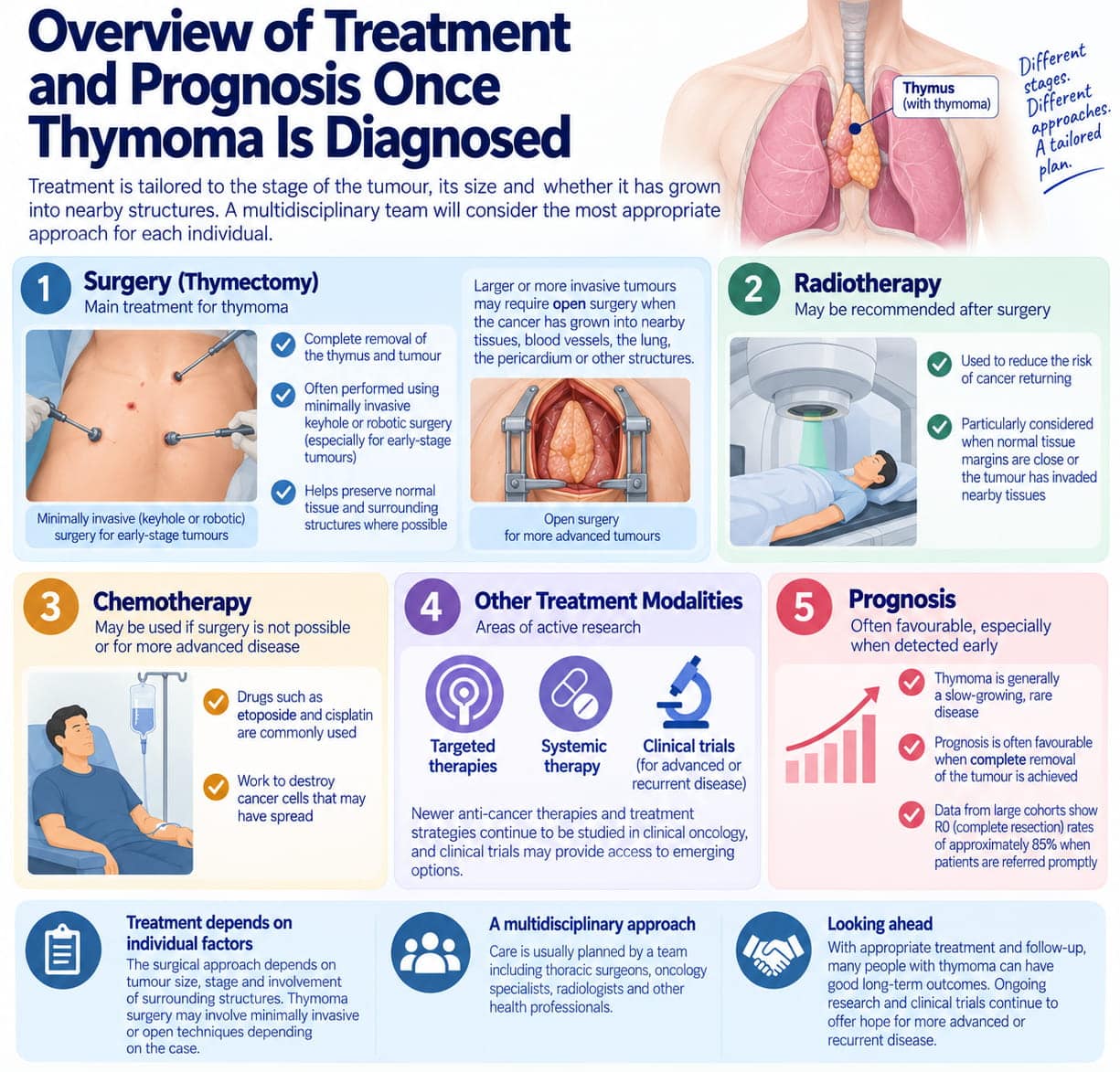
Surgery is the main treatment for thymoma, and thymectomy, the complete removal of the thymus and tumour, is the most common surgical approach.
This primary treatment can often be performed using minimally invasive keyhole or robotic surgery techniques in experienced cardiothoracic surgery centres, particularly for early-stage tumours, where normal cells and healthy tissue surrounding the gland can be preserved where appropriate.
Larger or more invasive tumours may require open surgery when the cancer has grown into nearby tissues, blood vessels, the lung, the pericardium, or other structures.
Radiotherapy (radiation therapy) may follow surgery to reduce the risk of cancer returning, particularly when normal tissue margins are close, or the tumour has invaded nearby tissues.
Chemotherapy may be used if surgery is not possible or for more advanced disease. Chemotherapy drugs can include etoposide and cisplatin, which work to destroy cancer cells that may have spread.
Additional treatment modalities, including targeted therapies and systemic therapy, remain areas of active research in clinical oncology.
Because thymoma is generally a slow-growing rare disease, prognosis is often favourable when the disease is detected early, and complete removal of the tumour is achieved. Data from large cohorts show complete resection, or R0 resection, rates of approximately 85% when patients are referred promptly.
The surgical approach depends on tumour size, stage, and involvement of surrounding structures, so that thymoma surgery may involve minimally invasive or open techniques depending on the individual case.
Patients with advanced or recurrent disease may also be considered for clinical trials, which can provide access to newer anti-cancer therapies and emerging cancer treatment strategies.
Not sure about your treatment options?
Mr Scarci provides expert consultations typically within one week of contact.
Living With Thymoma and Associated Autoimmune Diseases
Even after successful thymoma surgery, autoimmune diseases such as myasthenia gravis, pure red cell aplasia and other autoimmune diseases may persist and need long-term management by neurologists, immunologists or haematologists.
The immune system does not always reset after the tumour is removed, so complete removal of the thymoma does not necessarily mean that every associated autoimmune condition will disappear.
Regular follow-up appointments and periodic chest CT scans are important to monitor for recurrence, particularly during the first few years after surgery when the risk may be higher.
People diagnosed with thymoma may also experience emotional and practical challenges associated with having a rare condition, including anxiety about cancer returning and uncertainty about long-term health.
Multidisciplinary cancer care involving thoracic surgery, clinical oncology, neurology, immunology and specialist nursing helps coordinate treatment of both the tumour and associated paraneoplastic syndromes.
Patients under the care of specialists such as Mr Scarci may also receive tailored post-operative support, rehabilitation, and guidance on returning to normal activities, travel, and work.
Frequently Asked Questions About Early Thymoma Symptoms
Can thymoma cause symptoms before it is visible on a chest X-ray?
Yes. Very small thymomas may not appear on a standard chest X-ray but can still be detected by a more sensitive CT scan. Early immune-related symptoms such as myasthenia gravis can develop before the tumour is large enough to show on plain radiography. When normal cells form abnormally within the thymus, the mass may be too small to detect on X-ray but large enough to trigger an immune response.
Are early thymoma symptoms different from those of thymic carcinoma?
Early symptoms can overlap. Cough, chest pain and breathlessness are common to both. However, thymic carcinomas are rarer and tend to grow more aggressively, so symptoms may progress more quickly. Systemic signs such as significant weight loss and fatigue can appear earlier with thymic carcinoma compared with typical slow-growing thymoma. Thymic carcinomas are also more likely to metastasise to bones and liver, potentially causing symptoms at distant sites.
Could my autoimmune disease mean I already have a thymoma?
While conditions such as myasthenia gravis, pure red cell aplasia and other autoimmune diseases are more common in people with thymoma, most individuals with these conditions do not have a thymic tumour. However, lymph nodes and anterior mediastinal structures may need assessment. Your doctor may recommend a chest CT scan if the clinical picture raises concern, especially if you have unexplained findings on blood tests or symptoms involving multiple organ systems.
How quickly do early thymoma symptoms usually progress?
Thymomas often grow slowly over months or years, so early symptoms can be mild and gradually progressive. Any noticeable change in breathing, chest discomfort or muscle strength over weeks to months should be assessed rather than watched indefinitely. The rate of progression can vary depending on the histological type. A predominantly cortical thymoma, for example, may behave differently from a Type A tumour.
Who should I see if I am worried about possible thymoma symptoms?
Start with your GP for initial evaluation and imaging. If findings suggest a mediastinal mass, request referral to a specialist thoracic surgery or thoracic oncology service. A specialist thoracic surgeon can assess suspected thymic tumours and discuss minimally invasive treatment options, including robotic surgery where appropriate.
