

Discovering a painful lump under your rib cage can be worrying, especially when you are unsure whether it comes from the ribs, chest wall or an organ beneath them. Many lumps have a straightforward cause, such as a strained muscle, small hernia or benign growth, but a new or persistent swelling should still be properly assessed.
The location, size, texture and associated symptoms can provide important clues about the cause. Knowing which warning signs require prompt medical attention can also help you avoid unnecessary worry while making sure more serious conditions are not overlooked.
This guide explains the common causes of a lump under the rib cage, the warning signs to look for, how these lumps are diagnosed and when specialist assessment may be needed.
A painful lump under the rib cage is common and often benign, but it can occasionally signal serious problems involving the ribs, chest wall or abdominal organs. A lump under the rib cage can originate from superficial skin and fat tissue, from the rib bones or cartilage themselves, from the chest or abdominal wall muscles, or occasionally from deeper abdominal organs.
The most frequent causes include hernias, benign soft-tissue growths such as lipomas, inflamed cartilage, rib injuries, and, less commonly, enlarged organs or tumours in the right or left upper quadrant.
Red-flag symptoms that need urgent assessment include sudden severe pain, a rapidly enlarging or very hard lump, fever and feeling unwell, difficulty breathing, jaundice, or a pulsating lump in the abdomen.
Diagnosis usually requires a medical professional to perform a physical examination of the rib cage and abdomen. It may include blood tests and imaging such as ultrasound, CT scan or MRI.
What Does a Painful Lump Under the Rib Cage Mean?
A lump under the rib cage may arise from the chest wall, including bone, cartilage, muscle or skin, or from an underlying abdominal mass in the upper abdomen beneath the ribs. It can be caused by many things, and the location matters. Patients often first notice a swelling under the right rib cage or left side when pressing, twisting or breathing deeply. Some lumps appear only when standing or coughing, while others are noticed incidentally while showering or dressing.
Anatomically, the right upper quadrant of the abdomen contains the liver and gallbladder, while the left upper quadrant houses the spleen, part of the stomach and pancreas. The kidney sits deeper on each side. A lump’s location, texture, whether it feels soft or hard, whether it moves, and whether it changes with position or coughing all help a specialist narrow down the underlying cause.
Most lumps are not cancer. However, any new persistent swelling under the ribs should be properly assessed rather than self-diagnosed. Symptoms can sometimes overlap with pain around the ribs, which may arise from a wide range of chest wall and thoracic conditions.
Want a specialist opinion on your condition?
Get a clear answer from a surgeon who will be with you throughout.
Common Chest Wall Causes: Ribs, Cartilage and Muscles
The chest wall is a frequent source of painful lumps. These often relate to bone, cartilage, connective tissue or muscle rather than internal organs. They typically become more obvious with movement, deep breathing or after injury. They may be tender to touch but are not usually associated with systemic symptoms such as weight loss or night sweats.
A true chest wall lump can arise from several different tissues, so examination and appropriate imaging are important when swelling persists or changes. The causes below are generally non-cancerous. Malignancy is discussed later.
Costochondritis and Inflamed Rib Cartilage
Costochondritis is inflammation of the cartilage connecting ribs to the breastbone or along the costochondral junction. It causes sharp, localised pain that can sometimes feel like a lump, particularly when pressing on the ribs, coughing or taking a deep breath. However, there is no true growing mass on scans.
Typical triggers include minor chest trauma, repetitive strain from exercise, respiratory infections or prolonged coughing episodes.
Management may include:
Rest and avoidance of aggravating movements
Anti-inflammatory medication such as ibuprofen when appropriate
Physiotherapy
Local steroid injections in resistant cases
Costochondritis is uncomfortable but not dangerous and usually settles over weeks to months. Persistent symptoms may warrant a chest wall pain assessment, while costochondritis symptoms can help distinguish inflammation from other causes of rib and chest discomfort.
"After two years of being told my symptoms were anxiety, Mr Scarci identified the problem immediately. The surgery was straightforward and I was discharged after three days. I only wish I had found him sooner."
Rib Fractures, Bruises and Post-Traumatic Lumps
Direct chest trauma to the right or left ribs, from falls, sports injuries or road traffic accidents, can cause bruising, a haematoma or fractures that feel like a hard, painful lump. During healing, a callus of new bone forms, which can sometimes leave a firm ridge or bump along the rib cage.
Symptoms suggesting a fracture or serious internal injury include severe pain with breathing, shortness of breath, visible deformity or significant bruising spreading across the chest or abdomen. Initial assessment typically involves a chest X-ray or CT scan.
Most rib injuries are treated conservatively with pain control, breathing exercises and time, as outlined in this guide to broken rib treatment. Persistent painful bony lumps or deformity can sometimes require specialist thoracic assessment. At the same time, patients who are uncertain whether trauma has caused a fracture can review the common signs of broken ribs.
Muscle Strain and Intercostal Haematoma
Intercostal muscle strain is a common cause of localised aching or sharp pain along the rib cage, sometimes accompanied by subtle swelling after overuse, intense exercise or repeated coughing. Localised haematomas can result from physical trauma or heavy lifting, as small tears in muscle or blood vessels between the ribs may create a tender and sometimes bruised lump.
Management is generally straightforward, with rest, ice or heat, appropriate pain relief and gradual return to activity. These soft-tissue lumps often resolve within days to weeks, but ongoing symptoms should prompt review to rule out more serious causes.
Any history of significant injury, anticoagulant medication use or an unexplained large haematoma needs prompt medical attention because broken rib complications can include problems such as pneumothorax or flail chest.
Abdominal Wall Hernias Near the Rib Cage
Hernias are one of the most frequent causes of a visible lump between the rib cage and upper abdomen. A hernia occurs when a weakness in the abdominal wall allows fat or part of an internal structure to push through, creating a bulge.
Hernias can develop due to increased abdominal pressure from coughing, straining, or heavy lifting. They may also develop during pregnancy. Typical features include a soft lump that may be pushed back in, a burning or tugging sensation, and enlargement over time.
Hernias do not usually heal by themselves. Surgery is the definitive treatment for symptomatic hernias, and urgent assessment is necessary if the lump becomes suddenly painful, hard or irreducible, which can suggest strangulation.
Epigastric and Incisional Hernias
Epigastric hernias often cause lumps below the rib cage, appearing between the lower sternum and the belly button. They commonly contain fatty tissue, although bowel may occasionally become involved. This can create a risk of bowel obstruction or strangulation, presenting with severe abdominal pain, vomiting and distension.
Incisional hernias appear along previous surgical scars in the upper abdomen, particularly following operations involving the stomach, gallbladder or liver. Diagnosis is usually clinical but may be confirmed with ultrasound or CT scanning. Repair techniques include open and laparoscopic, or keyhole, surgery.
Not sure about your treatment options?
Mr Scarci provides expert consultations typically within one week of contact.
Benign Soft-Tissue Lumps Under the Rib Cage
Benign soft-tissue lumps are a common reason for painless or mildly painful swellings under the rib cage, often detected incidentally. These growths typically arise from fat, connective tissue, skin or blood vessels of the chest wall rather than internal organs.
Benign tumours can develop in various tissues of the chest wall. Although they are usually non-cancerous, any new or changing lump should be examined by a medical professional to exclude malignancy. Physical examination, combined with ultrasound or MRI, can often distinguish benign lipomas or cysts from lesions that require further investigation.
Mr Marco Scarci evaluates chest wall lumps as part of his wider expertise in thoracic and chest wall conditions.
Lipomas and Other Benign Fatty Tumours
Lipomas are soft-tissue tumours made of fatty tissue. They are usually slow-growing, soft, rubbery lumps beneath the skin and frequently occur on the chest wall, back, abdomen or shoulders. They are generally painless and mobile.
Most lipomas are small and do not require treatment unless they become painful, enlarge or cause cosmetic or functional concerns. Ultrasound or MRI may be used to confirm the diagnosis, especially if the lipoma lies close to ribs, nerves or a major blood vessel.
Surgical excision is usually a short procedure under local or general anaesthesia, and recurrence after complete removal is uncommon.
Cysts, Abscesses and Skin Lumps
Common skin and subcutaneous lesions under the rib cage include epidermoid cysts, which often feel firm and smooth and may have a small central opening. Cysts can form anywhere in the body, including the abdomen and chest wall.
Bacterial infection of a cyst or hair follicle can lead to an abscess, producing a hot, red and very tender lump.
Treatment options include:
Observation for small, non-infected cysts
Antibiotics and drainage for an abscess
Planned excision for recurrent or enlarging lesions
Any skin lump that bleeds, changes colour or develops irregular edges should be medically reviewed. Deeper chest wall abscesses should never be drained at home because they may connect with deeper structures or require imaging-guided treatment.
"I had rib pain for 6 years, was bounced around NHS without diagnosis. I was recommended to Mr Scarci — at consultation within minutes he knew what the problem was. Was offered surgery date the next week. 2 weeks post surgery I am almost back to full health."
Enlarged Organs and Abdominal Masses Under the Ribs
Some painful or firm lumps beneath the rib cage arise from enlarged organs rather than the chest wall. The liver and gallbladder sit beneath the right rib cage, while the spleen and parts of the stomach and pancreas lie beneath the left side.
These masses are often deeper and may be associated with symptoms such as fatigue, fever, weight loss or jaundice. Diagnosis requires careful examination and imaging and may involve collaboration between thoracic surgery, hepatobiliary surgery and gastroenterology.
Although serious conditions are possible, many cases of organ enlargement are caused by infections or benign disease that can be treated.
Right Upper Quadrant: Liver and Gallbladder
A firm, sometimes tender fullness under the right rib cage may reflect an enlarged liver, or hepatomegaly. Potential causes include fatty liver disease, viral hepatitis, heart failure, fluid accumulation or liver tumours.
Gallbladder disease, including cholecystitis or gallstones, can cause severe pain in the right upper quadrant that sometimes radiates towards the back or right shoulder. Associated symptoms requiring urgent assessment include fever, chills, jaundice, dark urine or pale stools.
Standard investigations include liver function blood tests, abdominal ultrasound, and CT or MRI if further detail is required. Treatment depends on the underlying cause and may involve antibiotics, endoscopic treatment or surgery.
Left Upper Quadrant: Spleen, Stomach and Pancreas
Splenomegaly is an enlarged spleen and may present as a palpable mass in the left upper quadrant. An enlarged spleen can sometimes be felt as a lump in the abdomen and may cause fullness, early satiety or discomfort during deep breathing.
Potential causes include liver disease, infections such as glandular fever, blood disorders, leukaemias and lymphomas. Imaging such as ultrasound or MRI can help confirm enlargement, while treatment depends on the underlying cause.
Masses involving the stomach or pancreas can also occur in this area, sometimes accompanied by indigestion, unexplained weight loss or changes in appetite. Persistent abdominal pain or a steadily enlarging mass warrants proper medical review and imaging.
Have a question about your diagnosis?
A consultation gives you a personalised plan — not a generic estimate.
When a Lump Under the Rib Cage Might Be Cancer
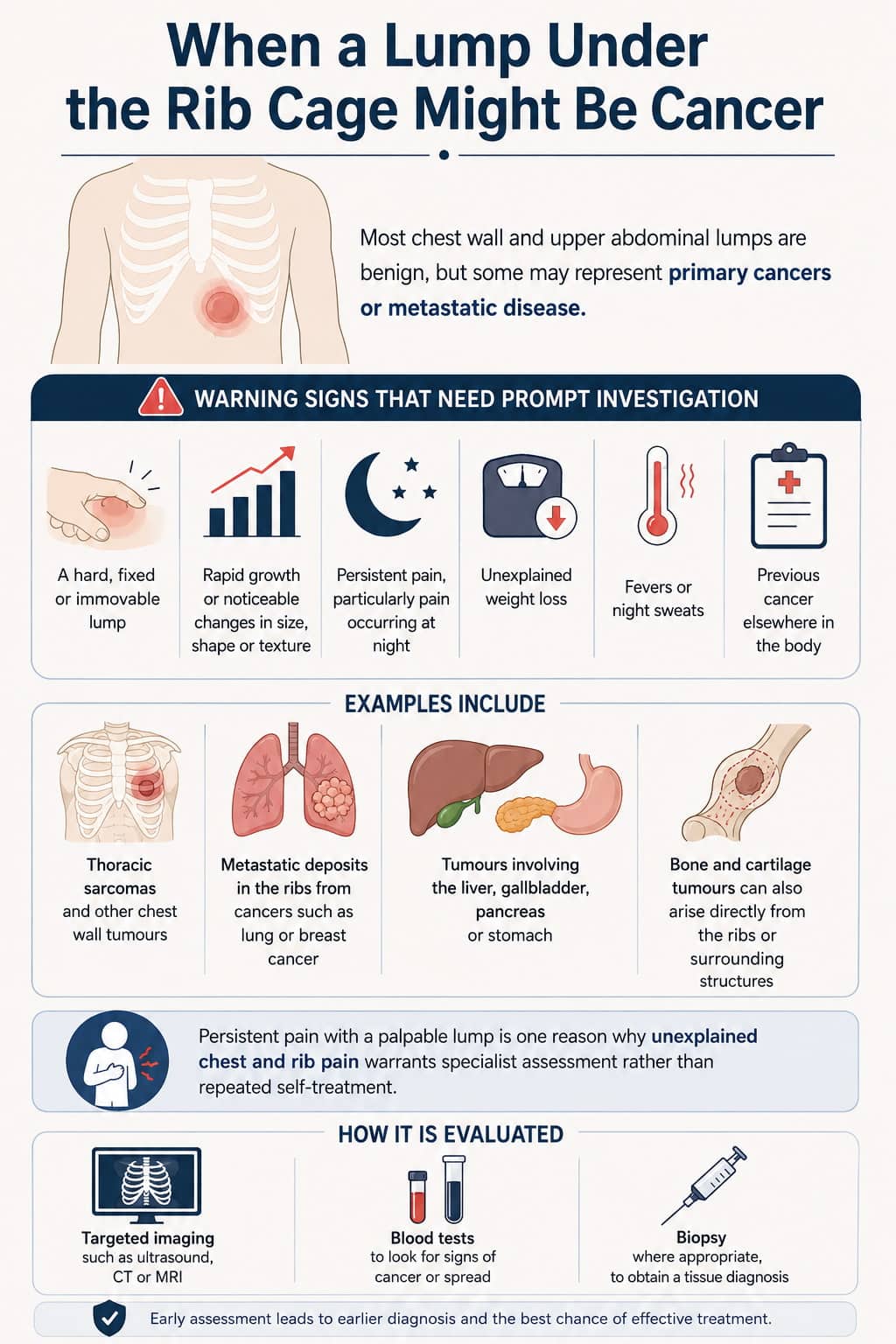
Most chest wall and upper abdominal lumps are benign, but some may represent primary cancers or metastatic disease.
Warning signs that may require more urgent investigation include:
A hard, fixed or immovable lump
Rapid growth or noticeable changes in size, shape or texture
Persistent pain, particularly pain occurring at night
Unexplained weight loss
Fevers or night sweats
Previous cancer elsewhere in the body
Examples include thoracic sarcomas and other chest wall tumours, metastatic deposits in the ribs from cancers such as lung or breast cancer, and tumours involving the liver, gallbladder, pancreas or stomach. Bone and cartilage tumours can also arise directly from the ribs or surrounding structures.
Persistent pain with a palpable lump is one reason why unexplained chest and rib pain warrants specialist assessment rather than repeated self-treatment. Evaluation typically involves targeted imaging, blood tests and, where appropriate, biopsy.
How Specialists Investigate a Painful Lump Under the Ribs
When a patient presents with a lump under the rib cage, the typical pathway involves:
Detailed history: Onset, chest trauma or injury, associated symptoms, smoking, alcohol use, previous cancers and recent infection.
Physical examination: Palpation of the chest wall and abdomen, checking the lump’s size, texture, mobility and relationship to breathing or movement.
Initial tests: Blood tests, chest X-ray and ultrasound of the right or left upper quadrant where appropriate.
Advanced imaging: CT scan or MRI of the thorax or abdomen if the lump is deep, fixed or suspicious.
Biopsy: If a chest wall mass is suspected, image-guided or surgical biopsy may be required.
CT and MRI can help define the relationship between a lump and the ribs, muscles, lungs and surrounding soft tissues. For lower-rib symptoms where standard investigations appear normal, conditions affecting the mobile ribs may also need consideration.
Persistent discomfort around the lowest ribs is discussed further in this guide to floating rib pain. A clear diagnosis is essential before deciding on observation, medication, minimally invasive surgery or more extensive treatment.
Want a specialist opinion on your condition?
Get a clear answer from a surgeon who will be with you throughout.
When to Seek Urgent or Emergency Medical Care
There is an important difference between routine assessment and red-flag symptoms requiring same-day or emergency care. Medical evaluation is recommended for lumps that persist, but certain features require immediate action.
Call 999 or attend A&E if you experience:
Sudden onset of severe chest or upper abdominal pain
A rapidly enlarging or extremely tender lump
Marked difficulty breathing or coughing up blood
Collapse, fainting or high fever with rigours
A pulsating abdominal lump
A pulsating abdominal mass can indicate an abdominal aortic aneurysm, which is a life-threatening emergency involving a major blood vessel. Conditions such as a strangulated hernia, perforated ulcer, severe liver or gallbladder infection, or traumatic chest injury can also be life-threatening and should not be ignored.
Do not drive yourself if you feel unwell, faint or light-headed. If the lump is stable, mildly uncomfortable and not accompanied by systemic symptoms, it is usually reasonable to arrange a prompt non-emergency consultation.
How Mr Marco Scarci Can Help

Mr Marco Scarci is a consultant thoracic surgeon based in London, experienced in diagnosing and treating chest wall, rib and lung conditions in both NHS and private settings. He specialises in minimally invasive thoracic surgery, including keyhole, VATS and robotic techniques where appropriate.
He provides face-to-face and virtual consultations for patients with painful lumps under the rib cage, helping distinguish chest wall problems from abdominal or organ-related causes. Where the cause falls within thoracic surgery, including chest wall masses, rib deformities, pleural disease or lung pathology, treatment can be tailored to the individual diagnosis.
If investigations reveal a condition outside thoracic surgery, such as liver, gallbladder or kidney disease, appropriate referral to another specialist can be arranged. For complex cases involving several areas of care, a multidisciplinary thoracic team can help coordinate assessment and treatment.
Looking After Yourself While You Wait for Assessment
While waiting for investigations, practical steps can help you monitor symptoms and avoid making discomfort worse:
Keep a symptom diary: Note pain levels, lump size, triggers, weight loss, fever or other changes.
Use simple pain relief: Over-the-counter pain medication may be appropriate for some people, alongside warm or cold packs.
Avoid heavy lifting: This is particularly important when a hernia or chest wall injury is suspected.
Do not repeatedly press the lump: Frequent manipulation can increase inflammation, bruising or tenderness.
Support your general health: Maintain a healthy weight, moderate alcohol intake and stop smoking where possible.
Attend all scheduled scans and follow-up appointments, and contact the clinic or seek medical advice sooner if symptoms significantly worsen or new red-flag signs develop.
Frequently Asked Questions
Is a painful lump under my right rib always related to the liver or gallbladder?
No. While the liver and gallbladder sit in the right upper quadrant and can cause deep-seated pain, many lumps under the right side of the rib cage are due to chest wall issues – rib injuries, inflamed cartilage, lipomas, or small hernias. Only a physical examination and imaging can reliably distinguish between chest wall and organ-related causes. Do not jump to conclusions about liver or gallbladder disease based solely on the location of the pain.
The lump moves when I press it and is softer when I lie down. Is that a good sign?
Lumps that change with position, coughing, or straining are often hernias or benign soft-tissue lesions, which tend to be less worrying than fixed, rock-hard masses. However, “good sign” does not mean “safe to ignore” – even small hernias can become trapped or strangulated. Seek urgent care if a previously soft, reducible lump suddenly becomes hard, extremely painful, or cannot be pushed back in.
Can anxiety or stress cause a lump under my rib cage?
Anxiety itself does not create a physical lump, but it can increase body awareness and muscle tension, making normal structures or minor strains feel more noticeable or painful. Any real, persistent swelling should still be examined to exclude a physical cause. Discuss both physical and emotional symptoms with your clinician, as addressing anxiety can improve overall symptom control.
Will I always need surgery for a lump under my ribs?
Not always. Treatment depends entirely on the underlying cause. Many lumps – such as small lipomas, muscle strains, or mild costochondritis – can be managed without surgery. Surgery is more likely for symptomatic hernias, suspicious or confirmed tumours, large painful lipomas, or structural rib problems affecting breathing or function. A specialist will discuss all options and explain benefits and risks before any procedure.
How quickly should I get a new lump under my rib cage checked?
Any new lump lasting longer than two to three weeks, or earlier if it is painful or enlarging, should be assessed by a medical professional. Seek urgent (same-day or emergency) review if there is severe pain, fever, breathing difficulty, jaundice, or a pulsating abdominal mass. If you are unsure whether your symptoms are urgent, contact NHS 111 for triage or arrange a prompt consultation with a medical professional.
