

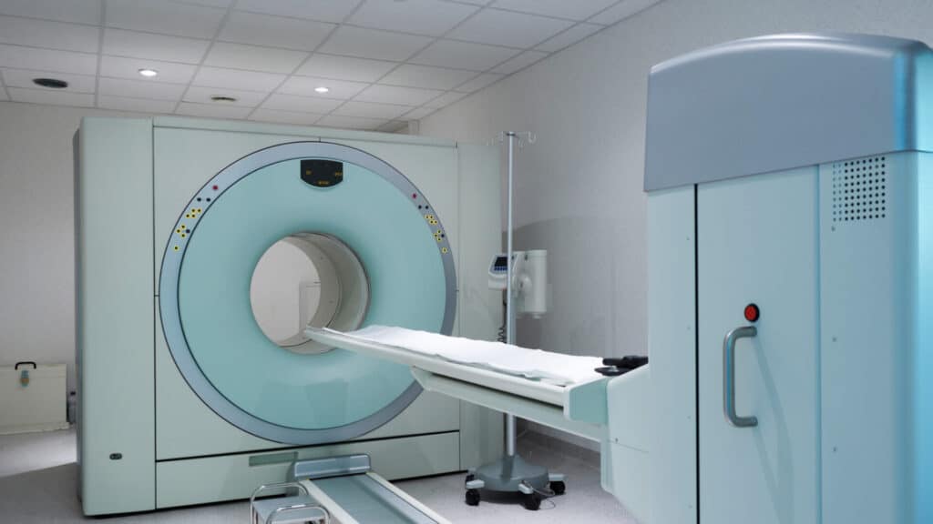
PET-CT is an important part of lung cancer staging because it combines detailed anatomical imaging with information about how metabolically active a tumour or lymph node is. For patients being considered for curative treatment, the scan can identify disease that is not obvious on CT alone, clarify whether suspicious lymph nodes need biopsy, and help determine whether surgery, radiotherapy, systemic therapy, or a combination is most appropriate.
This guide explains how PET-CT works, why it is used before lung cancer surgery, what its limitations are, and what patients can expect before and after the scan.
PET-CT combines PET and CT imaging to assess both the location and metabolic activity of lung cancer.
NICE recommends PET-CT before treatment for patients with lung cancer who may be suitable for curative-intent treatment.
PET-CT can identify unsuspected lymph node or distant metastatic disease that changes the treatment plan and may prevent non-beneficial surgery.
PET-CT does not replace biopsy and can produce false-positive and false-negative findings.
Accurate staging helps determine whether surgery, radiotherapy, chemotherapy, targeted treatment or another approach is most appropriate.
Symptoms, Early Detection and Accurate Lung Cancer Staging
Lung cancer remains a major cause of cancer-related mortality in the UK and worldwide. GLOBOCAN 2022 recorded approximately 50,700 new lung cancer cases in the UK across men and women. Earlier diagnosis can create more treatment options, including potentially curative surgery for appropriately selected patients, while advanced or metastatic lung cancer generally requires a different treatment approach.
Common symptoms that can prompt investigation include persistent coughing, shortness of breath, breathing difficulty, unexplained weight loss, reduced appetite, lethargy, weakness and reduced exercise tolerance. Some people have few or no symptoms when lung cancer is first identified, while high-risk individuals may enter lung cancer screening pathways where abnormalities can be detected before symptoms develop.
Types of Lung Cancer
Non-small cell lung cancer (NSCLC) accounts for approximately 85% of lung cancers and includes adenocarcinoma, squamous cell carcinoma and several less common subtypes. NSCLC is the type most commonly considered for surgical treatment when the disease is sufficiently localised.
Small cell lung cancer (SCLC) behaves differently and is more often treated with systemic therapy and radiotherapy, although staging remains essential when deciding between limited- and extensive-stage treatment approaches. Lung cancer may also be primary, originating in the lung itself, or metastatic, where cancer from another organ has spread to the lung.
Why Imaging Matters
Lung cancer prognosis and treatment depend heavily on tumour size, local invasion, lymph node involvement and distant spread. This is why lung cancer staging is so important: the TNM system evaluates the primary tumour, regional lymph nodes and distant metastases to establish disease stage.
Accurate staging determines whether surgery, radiotherapy, systemic treatment or a combination offers the most appropriate chance of cure or long-term disease control. For patients being considered for lung cancer surgery, PET-CT is interpreted alongside diagnostic CT imaging and other investigations before an operation is planned.
Want a specialist opinion on your condition?
Get a clear answer from a surgeon who will be with you throughout.
What Is a PET-CT Scan in Lung Cancer?
A PET-CT scan combines positron emission tomography (PET) with computed tomography (CT) during the same examination. It brings together metabolic information from PET with the detailed anatomical information provided by CT.
The PET component involves injecting a small amount of radioactive tracer, most commonly 18F-fluorodeoxyglucose (FDG), into a vein. FDG is a glucose analogue, and many cancer cells consume glucose at a higher rate than surrounding normal tissue, meaning areas of increased FDG uptake may appear more prominent on PET imaging.
The CT component provides high-resolution anatomical images showing the location, size and shape of lung tumours, lymph nodes and other structures. When the two scans are combined, clinicians can see both where a lesion is and how metabolically active it appears.
A standard lung cancer PET-CT commonly covers much of the body from the skull base to the upper thighs. This allows the primary lung tumour, regional lymph nodes and common sites of distant metastasis such as the adrenal glands, liver and bones to be assessed in a single examination.
"Dr. Marco Scarci is a deeply knowledgeable, open-minded, and empathetic surgeon. He listens to details and solves problems to truly deliver the most optimal results. As a medical professional myself, I can tell you that finding a specialist like Dr. Scarci is very rare, and it is a privilege to have him in my corner."
How PET-CT Fits Into Diagnosing Lung Cancer
Lung cancer diagnosis and staging usually follow a structured pathway rather than relying on one test. Imaging, tissue sampling and assessment of the patient’s general health are brought together before treatment is recommended.
Initial Imaging and Suspicion
A chest X-ray may be the first investigation when symptoms raise concern about lung disease. If a pulmonary mass or suspicious nodule is identified, a contrast-enhanced CT scan is usually performed to provide more detailed anatomical information.
When CT findings suggest lung cancer that could potentially be treated with curative intent, PET-CT is used to refine staging. CT remains particularly important for examining the lung itself, while MRI has a more specialised role where greater soft-tissue detail is required, as explained in the comparison of CT and MRI for thoracic imaging.
Biopsy and Tissue Confirmation
PET-CT alone cannot provide a definitive histological diagnosis or establish the exact cancer subtype. Tissue or cell sampling is generally required to determine the diagnosis and guide treatment.
Biopsy may be performed through bronchoscopy, endobronchial ultrasound-guided biopsy, CT-guided biopsy or surgical techniques. PET-CT can help identify which lesion or lymph node is most metabolically active and potentially the most useful target for obtaining diagnostic tissue.
Multidisciplinary Team Review
After imaging and biopsy, results are usually discussed at a multidisciplinary team (MDT) meeting. CT, PET-CT, biopsy findings, lung function and overall patient fitness are considered together to recommend surgery, radiotherapy, systemic therapy or a combination.
Mr Marco Scarci is a London-based consultant thoracic surgeon who reviews PET-CT findings alongside CT imaging, biopsy results, lung function and MDT recommendations when assessing whether minimally invasive or more complex thoracic surgery is appropriate.
Have a question about your diagnosis?
A consultation gives you a personalised plan — not a generic estimate.
Understanding Lung Cancer Staging and Why PET-CT Is Central
Lung cancer staging uses the TNM system. T, or tumour, describes the size and position of the primary cancer and whether it has invaded nearby structures; N, or nodes, describes whether cancer has spread to regional lymph nodes; and M, or metastasis, describes whether cancer has spread to distant parts of the body.
These categories are combined to assign an overall stage:
Stage | General Meaning | Typical Treatment Direction |
I | Localised tumour without nodal or distant spread | Surgery or another radical local treatment |
II | Larger tumour and/or limited nodal involvement | Surgery in suitable patients, often with additional systemic treatment |
III | Locally advanced disease and/or more extensive nodal involvement | Multimodality treatment; surgery in selected cases |
IV | Distant metastatic disease | Systemic treatment and other therapies based on disease biology and symptoms |
Treatment is more nuanced than the table alone can show. Tumour biology, molecular testing, performance status, lung function and patient preferences also influence which treatment options are appropriate.
PET-CT contributes particularly to the N and M components by detecting metabolically active lymph nodes and distant lesions that may not be obvious on anatomical imaging alone. Accurate staging can therefore prevent patients from undergoing major surgery when cancer has already spread beyond the point at which resection would provide the intended curative benefit.
"After multiple failed consultations elsewhere, Mr Scarci identified exactly what was wrong and operated within two weeks. Recovery was smooth and I'm back to normal life."
CT Scan vs PET-CT: What Each Test Shows
CT and PET-CT answer different clinical questions, which is why both are needed during lung cancer assessment.
Feature | CT Scan | PET-CT |
Main information | Anatomical size, shape and location | Anatomy plus metabolic activity |
Lymph nodes | Suspicion largely based on size and appearance | Detects increased FDG uptake |
Distant metastases | Detects visible anatomical lesions | Can identify metabolically active disease |
Scar vs active tumour | May be difficult to distinguish | Metabolic activity can add useful information |
Whole-body staging | Usually targeted anatomical coverage | Broader staging coverage in one examination |
A CT scan provides detailed images of tumour size, location and relationships with surrounding structures, but lymph node size alone is an imperfect marker of cancer involvement. Small nodes can contain metastatic disease, while enlarged nodes may simply be reactive or inflamed.
PET-CT adds metabolic information that can identify suspicious disease not apparent from size alone. This complementary relationship between anatomical and functional imaging is one reason CT and PET-CT are interpreted together rather than as competing investigations.
Not sure about your treatment options?
Mr Scarci provides expert consultations typically within one week of contact.
What Lung Cancer Looks Like on PET-CT
On a PET-CT scan, areas of greater uptake of the radioactive tracer appear as bright “hot spots” overlaid on the grey-scale CT images. The primary lung tumour may show increased FDG uptake, appearing as a prominent focus within the lung parenchyma. Involved lymph nodes, particularly hilar and mediastinal stations, may also show increased uptake when they harbour metastatic disease.
Radiologists and surgeons use a semi-quantitative measure called the standardised uptake value (SUV), specifically SUVmax, to gauge how metabolically active a lesion is. Higher SUV values can increase suspicion of malignant disease, but increased activity alone is not specific for malignancy. PET/CT can help distinguish suspicious from less suspicious nodules, but inflammatory or infectious conditions can also cause increased FDG uptake.
PET-CT highlights areas suspicious for cancer spread across multiple common metastatic sites, including mediastinal and hilar lymph nodes, adrenal glands, bones, and liver. PET scans can identify metabolically active lymph nodes that appear normal in size on CT, making them particularly valuable for accurate staging.
However, not all cancers show strong FDG uptake. Some low-grade adenocarcinomas, carcinoid tumours, and very slowly growing lesions may display minimal or absent uptake, meaning a negative PET scan does not always rule out malignancy. This is an important consideration when interpreting PET results alongside CT findings and clinical context.
Diagnostic Accuracy of PET-CT in Lung Cancer
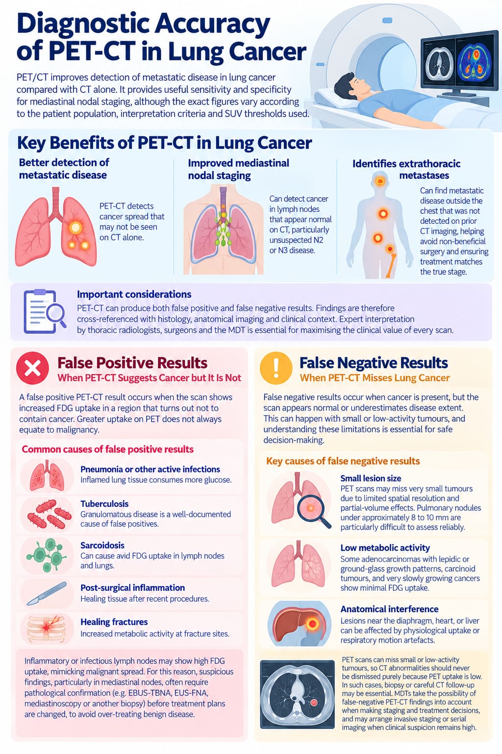
PET/CT improves detection of metastatic disease in lung cancer compared with CT alone. Studies and meta-analyses show that PET-CT provides useful sensitivity and specificity for mediastinal nodal staging, although the exact figures vary according to the patient population, interpretation criteria and SUV thresholds used.
PET scans can detect cancer spread to lymph nodes that appear entirely normal on CT, and this capability is particularly important for identifying unsuspected N2 or N3 mediastinal nodal disease that could change the treatment plan from surgery to chemoradiotherapy or systemic therapy.
PET-CT can also identify extrathoracic metastatic disease that was not detected on prior CT imaging. Identifying these cases avoids non-beneficial surgical interventions and helps ensure treatment matches the true stage of the disease.
Despite being highly useful, PET-CT still generates both false positive and false negative results. This is why PET results are cross-referenced with histology, anatomical imaging and clinical context. Expert interpretation by thoracic radiologists, surgeons and the MDT is important for maximising the clinical value of every scan.
Want a specialist opinion on your condition?
Get a clear answer from a surgeon who will be with you throughout.
False Positive Results: When PET-CT Suggests Cancer but It Is Not
A false positive PET-CT result occurs when the scan shows increased FDG uptake in a region that turns out not to contain cancer. Greater uptake on PET does not always equate to malignancy.
Common causes of false positive results include:
Pneumonia or other active infections: Inflamed lung tissue consumes more glucose
Tuberculosis: Granulomatous disease is a well-documented cause of false positives
Sarcoidosis: Can cause avid FDG uptake in lymph nodes and lungs
Post-surgical inflammation: Healing tissue after recent procedures
Healing fractures: Increased metabolic activity at fracture sites
PET scans can therefore yield false-positive results due to inflammation. Inflammatory or infectious lymph nodes may show high FDG uptake, mimicking malignant nodal spread on lung PET scan images.
This is precisely why suspicious PET-CT findings, particularly in mediastinal nodes, often require pathological confirmation through EBUS-TBNA, EUS-FNA, mediastinoscopy, or another appropriate biopsy procedure before treatment plans are changed. The aim is to balance the risk of over-treating benign disease against the risk of missing true cancer spread.
False Negative Results: When PET-CT Misses Lung Cancer
False negative results occur when cancer is present, but the scan appears normal or underestimates disease extent. False-negative results can occur with small or low-activity tumours, and understanding these limitations is essential for safe decision-making.
Key causes of false negatives include:
Small lesion size: PET scans may miss very small tumours because of limited spatial resolution and partial-volume effects. Pulmonary nodules under approximately 8 to 10 mm are particularly difficult to assess reliably.
Low metabolic activity: Some adenocarcinomas with lepidic or ground-glass growth patterns, carcinoid tumours, and very slowly growing cancers show minimal FDG uptake.
Anatomical interference: Lesions near the diaphragm, heart, or liver can be affected by physiological uptake or respiratory motion artefacts.
PET scans can miss small or low-activity tumours, which means CT abnormalities should never be dismissed purely because PET uptake is low. In such cases, biopsy or careful CT follow-up may remain essential for further investigation.
MDTs routinely account for the possibility of false-negative PET-CT findings when making lung cancer staging and treatment decisions. When clinical suspicion remains high despite a negative PET scan, invasive staging or serial imaging may be arranged to ensure significant disease is not missed.
Have a question about your diagnosis?
A consultation gives you a personalised plan — not a generic estimate.
Why PET-CT Is Essential Before Lung Cancer Surgery
Accurate lung cancer staging with PET-CT is crucial before offering curative-intent surgery such as lobectomy or segmentectomy, particularly via minimally invasive keyhole approaches. The goal is straightforward: ensure that patients only undergo lung cancer surgery and related procedures when there is a realistic chance of oncological benefit.
PET-CT can reveal previously unsuspected N2 mediastinal nodal disease or M1 distant metastases, which may change the plan from surgery to combined chemoradiotherapy, systemic therapy, or another multimodality approach. Studies have shown that PET-based staging can change management in a meaningful proportion of patients, with the effect varying according to the population studied.
Consider this representative scenario: a patient has a CT scan showing a 3.5 cm peripheral lung tumour with no enlarged lymph nodes. Based on CT alone, the surgeon plans a VATS lobectomy. PET-CT then reveals FDG-avid uptake in a mediastinal station 4 lymph node with an SUV of 3.5. EBUS biopsy confirms N2 disease, shifting the stage from I to IIIA.
The treatment plan can then be reconsidered before a major operation is performed. By avoiding non-beneficial thoracotomy or VATS in patients already upstaged by PET-CT, unnecessary surgical complications and recovery can be avoided.
How PET-CT Guides Surgical Planning for Mr Marco Scarci
Mr Scarci’s typical process begins with a thorough review of PET-CT results alongside CT imaging. The pattern of PET uptake in the primary lung tumour and surrounding lymph nodes directly influences the surgical approach, whether that is robotic lung cancer surgery, VATS lobectomy, segmentectomy, or wedge resection.
When PET-CT shows FDG-avid mediastinal lymph nodes, Mr Scarci may recommend additional invasive staging procedures such as EBUS or mediastinoscopy before committing to surgery. This step helps ensure that patients with confirmed N2 or N3 disease receive appropriate oncological treatment rather than proceeding directly to an operation that may not address all of their disease.
PET-CT also helps identify which lymph node stations may need to be sampled or dissected during surgery, supporting an oncologically complete operation. Accurate preoperative staging allows better counselling of patients about expected outcomes, risks, and the likelihood of needing additional treatment after surgery.
Both private and NHS patients treated by Mr Scarci in London hospitals can benefit from modern PET scanner technology and multidisciplinary interpretation, ensuring that each treatment plan is based on comprehensive staging information.
Not sure about your treatment options?
Mr Scarci provides expert consultations typically within one week of contact.
When Is PET-CT Ordered in the Lung Cancer Journey?
Doctors typically order a lung PET scan at several key time points:
Initial staging – after a suspicious CT scan suggests potentially operable lung cancer
Restaging – after initial treatment when reassessment is clinically indicated
Follow-up – if recurrence is suspected based on symptoms or surveillance imaging
For potentially operable NSCLC, PET-CT is requested soon after the diagnostic CT scan and before radical treatment is scheduled. If there is a significant delay before treatment, staging can become outdated, and repeat imaging may be needed.
PET-CT is less commonly used in small cell lung cancer for initial staging but can help differentiate limited disease from extensive disease in selected cases, particularly those being considered for radical treatment.
From a patient’s perspective, the scan usually takes place during the staging process after the initial CT has raised concern and before surgery is confirmed. The scan itself is performed as an outpatient visit and does not normally require an overnight stay.
Preparing for a Lung PET-CT Scan
Fasting and Water
Do not eat for 4 to 6 hours before the scan, unless your imaging department provides different instructions. This helps keep background glucose and insulin levels suitable so the radioactive tracer can be interpreted accurately.
You can usually drink plain water before the PET scan. Staying hydrated is generally encouraged.
Blood Sugar and Medications
Blood glucose is checked or managed according to the imaging centre’s protocol because elevated glucose can interfere with FDG uptake and scan quality. Patients with diabetes should discuss medication, insulin and meal timing with the imaging team in advance rather than changing their routine independently.
Tell the imaging team about diabetes, pregnancy, breastfeeding, and all current medications before the examination.
What to Expect During the Scan
Avoid strenuous exercise for around 24 hours before the scan if instructed, as muscle uptake of FDG can interfere with image quality. On the day itself, a radiographer places an intravenous cannula and injects the FDG tracer. You may feel a sharp sting during the injection, but the PET scan itself is painless.
After an uptake period while the tracer distributes throughout the body, you lie still in the PET scanner while images are acquired. The imaging portion commonly takes around 20 to 30 minutes, although the full appointment is longer because of preparation and tracer uptake time.
Radiation exposure from PET-CT is higher than from some individual imaging examinations, but when the scan is appropriately indicated for suspected or confirmed lung cancer, the clinical staging information generally outweighs the small radiation risk.
Want a specialist opinion on your condition?
Get a clear answer from a surgeon who will be with you throughout.
What to Expect From PET-CT Results and Next Steps
PET-CT reports are typically available after specialist interpretation, with timing varying between hospitals and imaging centres. In UK lung cancer pathways, results are commonly discussed at an MDT meeting so that imaging findings can be considered alongside pathology, lung function, and the patient’s overall condition.
The PET-CT findings are combined with lung cancer staging, biopsy results, lung function data, and overall fitness to recommend the next step: surgery, chemoradiotherapy, stereotactic radiotherapy, systemic therapy, or further investigation.
Patients should feel empowered to ask for a detailed explanation of their lung cancer staging and PET-CT report during consultation with a thoracic surgeon or oncologist. Understanding what each finding means, and what it does not mean, helps support informed decision-making.
Where PET-CT and biopsy results do not agree, additional tests such as repeat biopsy, EBUS, mediastinoscopy, or repeat imaging may be arranged.
Why Choose a Specialist Thoracic Surgeon for PET-CT-Guided Care
Reviewing PET-CT findings in the context of surgical anatomy, pathology, lung function, and overall fitness is an important part of treatment planning. Mr Marco Scarci brings specialist thoracic surgical experience to interpreting PET-CT alongside clinical findings when determining whether surgery is appropriate and which approach may be most suitable.
Mr Scarci’s focus on minimally invasive keyhole surgery, including VATS and robotic techniques, means accurate staging is especially important. These procedures can provide important recovery benefits in appropriate patients.
Personalised consultations review PET-CT, CT imaging, biopsy findings, and lung function to tailor both surgery and aftercare. International and UK-based patients may also seek specialist review when staging or treatment recommendations are unclear.
If you are uncertain about your lung cancer staging or proposed treatment, a second opinion consultation can provide another review of PET-CT and other investigations before a major treatment decision is made.
Frequently Asked Questions
Is a PET-CT scan always necessary before lung cancer surgery?
PET-CT is usually recommended before major surgery for potentially curable non-small cell lung cancer. Rare exceptions may apply, such as very small, low-risk nodules. Your MDT can explain whether it is needed in your case.
How long is PET-CT staging valid before surgery or treatment?
PET-CT results are usually considered current for a few weeks to a couple of months if symptoms remain unchanged. Repeat imaging may be needed after a longer delay or if new symptoms develop.
Does PET-CT replace the need for a lung biopsy?
No. PET-CT helps show the location and spread of suspected cancer, but a biopsy is usually needed to confirm the cancer type and guide treatment.
How risky is the radiation from a PET-CT scan?
PET-CT uses more radiation than a standard chest CT, but the exposure is within accepted medical limits. For suspected lung cancer, its staging information usually outweighs the small long-term radiation risk. Tell your imaging team if you are pregnant or may be pregnant.
Can I get a second opinion on my PET-CT before committing to surgery?
Yes. You can seek a second opinion before major lung surgery. Mr Marco Scarci can review your PET-CT, CT, and biopsy results remotely or in person and discuss your options.
