

A CT scan may reveal a “spot” or “shadow” on the lung, often referred to as a lung nodule. While most lung nodules are benign, some may be an early sign of lung cancer or require monitoring to rule it out.
Knowing what a lung nodule is and what follow-up may involve can help patients and families feel more informed and prepared.
Most lung nodules are benign. Risk depends on nodule size, appearance, growth, smoking history and other personal factors.
A pulmonary nodule is usually smaller than 3 cm. Larger nodules and those that grow over time carry a higher lung cancer probability.
CT scan follow-up is often all that is needed. PET CT, needle biopsy or surgery may be considered for more suspicious nodules.
Small pulmonary nodules are common and often result from previous infection, inflammation or scarring.
What Is a Lung Nodule and Should You Be Worried?
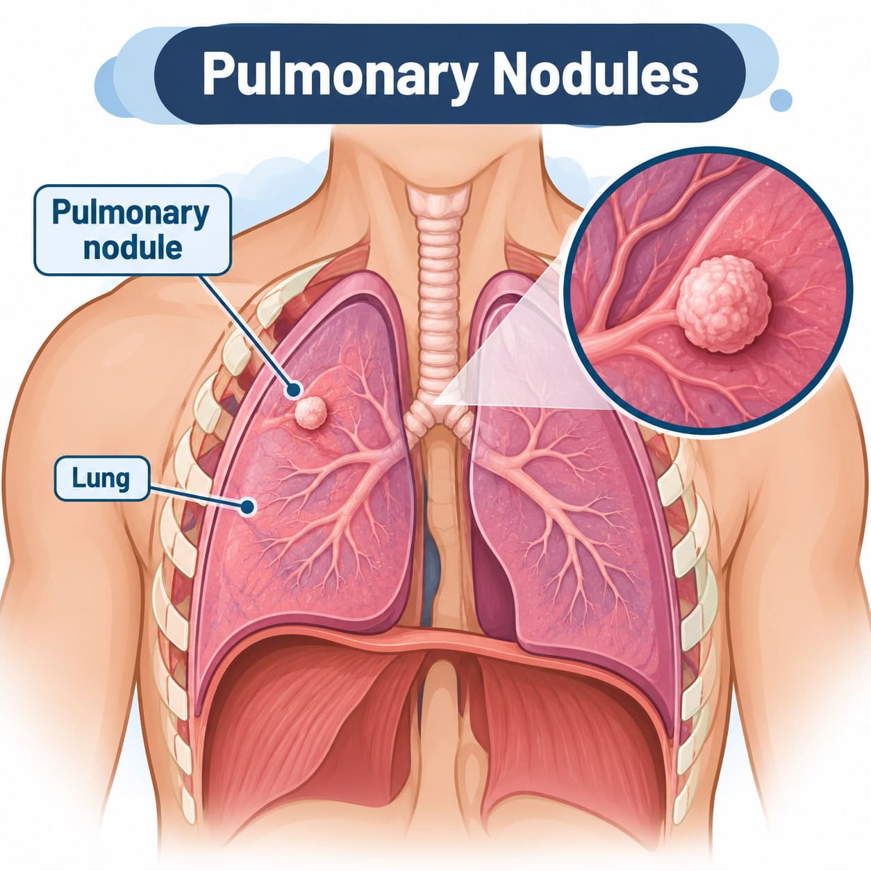
A lung nodule, also called a pulmonary nodule, is a small rounded area seen on a chest scan. Nodules are generally under 3 cm. Larger findings are called masses and need urgent assessment because their risk of cancer is higher.
Most nodules cause no symptoms and are found incidentally during a CT scan for another reason, such as cough, chest pain or a pre-operative check. Incidental pulmonary nodules detected on CT are also common in lung cancer screening programmes for high-risk individuals, including eligible current and former smokers.
The British Thoracic Society and Fleischner Society provide guidance to help doctors assess nodule risk and plan appropriate follow-up. A consultation with a thoracic specialist like Mr Marco Scarci can help determine whether a nodule needs further investigation, monitoring or treatment.
Can a Lung Nodule Turn into Cancer? The Direct Answer
Some lung nodules are cancerous when first detected. Others are benign and never change. A small number represent early abnormal cells that can develop into lung cancer over time.
Most small nodules are benign. A nodule that remains unchanged on CT scans for two years is very unlikely to be an active cancer, although ground-glass opacity and other subsolid nodules may need longer monitoring.
Features that increase lung cancer probability include:
Nodule size: Larger nodules have a greater risk of malignancy
Appearance: Spiculated edges, part-solid nodules and ground-glass opacity can be more suspicious
Growth: Increase in size or volume on serial CT images is an important warning sign
Personal risk: Older age, smoking history, family history and underlying lung disease increase risk
Early detection matters. When lung cancer is diagnosed at an early stage, treatment can often be curative.
How Common Are Lung Nodules and How Often Are They Cancerous?
Lung nodules are common, particularly in older people and those with a smoking history. Their malignancy status depends on the setting, nodule size and individual risk.
Setting | Typical Malignancy Rate |
Low-dose CT screening (e.g. National Lung Screening Trial, NELSON trial) | 3–5% |
Symptomatic patients referred to a lung cancer clinic | 40–60% |
Very small nodules (<6 mm) in low-risk adults | <1% |
The National Lung Screening Trial (NLST) enrolled 53,439 participants for lung cancer screening and demonstrated that low-dose CT screening can reduce lung cancer mortality. However, 96.4% of positive LDCT screens were false positives in that trial, meaning most flagged nodules turned out to be benign. This illustrates why guidelines aim to avoid unnecessary invasive procedures while still detecting lung cancer early.
Lung cancer probability rises steeply with nodule size: roughly 6% for nodules 5–10 mm, and up to 64% for nodules over 20 mm based on published data. The most common causes of benign nodules include old granulomas from previous infections, lymph nodes, and small areas of scarring.
Common benign causes include previous infection, inflammation, small lymph nodes and scar tissue. This is why a structured workup is preferable to assuming that every nodule is cancer.
Not sure about your treatment options?
Mr Scarci provides expert consultations typically within one week of contact.
Benign vs Malignant Lung Nodules: What Makes Them Different?
Radiologists use a qualitative assessment of CT images to estimate whether a nodule is more likely benign or malignant. No single sign provides a diagnosis.
Features suggesting a benign nodule:
Small nodule size
Smooth, well-defined edges
Central or diffuse calcification
Fat within the nodule
Stability on follow-up imaging
Features raising concern for a malignant nodule:
Growth on serial scans
Irregular or spiculated borders
Part-solid appearance or ground-glass opacity with an enlarging solid area
Upper lobe location
Pleural indentation or changes in nearby blood vessels
Infection and inflammation can mimic cancer, even on a PET scan. A tissue sample through biopsy or surgery may be needed to determine whether a suspicious nodule is cancerous.
The Role of Nodule Size: Why Millimetres Matter
Nodule size is a major factor in clinical practice, but clinicians consider it alongside appearance, growth and the patient’s condition.
Size Category | Approximate Risk | Usual Action |
<4 mm | Very low (<1%) in low-risk adults | Often no follow-up needed |
4–6 mm | Low (~1–2%) | CT follow-up in selected patients |
6–8 mm | Intermediate (~6%) | Careful surveillance with repeat CT |
8–30 mm | Higher (10–64%) | PET CT, biopsy, or surgery considered |
>30 mm | High (treated as suspicious mass) | Urgent investigation |
Nodules 4–12 mm are often classified as indeterminate pulmonary nodules, meaning they sit in a grey zone where imaging alone cannot confirm or exclude cancer. Nodules larger than 8 mm are at greater risk of malignancy than smaller nodules, and larger nodules are more likely to be diagnosed as lung cancer.
Lung cancer probability varies with nodule size and characteristics. Not only absolute size, but also growth over time is critical. Volume doubling, where the nodule’s volume doubles over a measurable period, is a strong signal of possible malignancy. Nodules less than 8 mm generally require CT follow-up, while those 8–30 mm may need biopsy or PET CT for further evaluation.
Small nodules under 8 mm are usually monitored with CT. Larger nodules, or nodules that grow, are more likely to require PET CT or biopsy.
Risk calculators, including the Brock model, combine nodule size, upper lobe location, smoking status, age and CT features to estimate lung cancer probability. They support, rather than replace, clinical judgement.
Have a question about your diagnosis?
A consultation gives you a personalised plan — not a generic estimate.
Growth and Change Over Time: How Fast Can a Lung Nodule Turn Malignant?
Growth is one of the most useful indicators of malignancy. Doctors compare current and previous CT images to determine whether nodules are stable, slowly growing or enlarging more quickly.
Many lung cancers increase in volume over months or years. Solid nodules that are unchanged over two years are usually benign. Subsolid nodules can grow more slowly and may need follow-up for up to five years.
Sudden change from a tiny, stable small nodule to advanced cancer is unusual. Where cancer develops, progressive growth is usually visible on scans before symptoms occur.
Who Is at Higher Risk? Patient Factors That Influence Malignancy
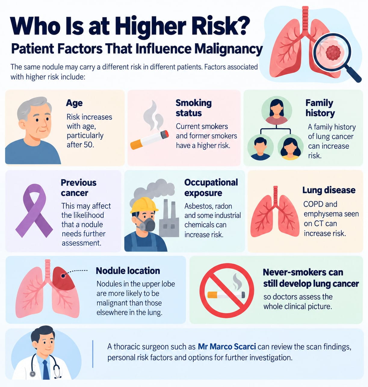
The same nodule may carry a different risk in different patients. Factors associated with higher risk include:
Age: Risk increases with age, particularly after 50
Smoking status: Current smokers and former smokers have a higher risk
Family history: A family history of lung cancer can increase risk
Previous cancer: This may affect the likelihood that a nodule needs further assessment
Occupational exposure: Asbestos, radon and some industrial chemicals can increase risk
Lung disease: COPD and emphysema seen on CT can increase risk
Nodules in the upper lobe are more likely to be malignant than those elsewhere in the lung. Never-smokers can still develop lung cancer, so doctors assess the whole clinical picture.
A thoracic surgeon such as Mr Marco Scarci can review the scan findings, personal risk factors and options for further investigation.
How Are Lung Nodules Found? Chest X‑ray vs CT Scan
Small pulmonary nodules may not appear on a chest X-ray. CT scans provide much more detail and can identify very small nodules.
Low-dose CT screening is used to identify lung cancer earlier in high-risk individuals. Evidence from the National Lung Screening Trial and NELSON trial supports low-dose CT screening as a way to reduce lung cancer mortality.
Nodules may also be found during emergency imaging, pre-operative assessment or CT for unrelated symptoms. Consistent scan technique, including slice thickness and reconstruction algorithm, helps doctors compare nodule size accurately over time.
CT is usually the first choice for assessing pulmonary nodules, while MRI may be used when further imaging is needed, or CT is not suitable.
How Do Doctors Decide If a Nodule Is Cancerous? Imaging Features and Risk Calculators
Doctors consider:
Nodule size, shape, density and location
Whether the nodule is solid, part-solid or ground-glass
Growth on follow-up CT scans
Smoking status, age, family history and other risk factors
PET CT findings where appropriate
The Fleischner Society, British Thoracic Society and Lung-RADS systems provide follow-up recommendations based on risk category. The Brock model estimates the chance of cancer, while the Herder model adds PET scan results.
Advanced image analysis from biomedical engineering research may improve discriminatory ability, but it is not yet standard in everyday care. Doctors use these tools alongside their clinical assessment and, when necessary, may recommend a lung biopsy to determine whether a nodule is cancerous.
The Role of PET CT: Metabolic Clues to Malignancy
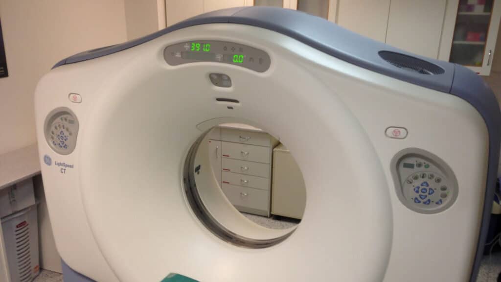
PET CT combines CT anatomy with a PET scan that measures metabolic activity. Cancerous nodules often use more glucose and may appear as a ‘hot spot’.
PET CT is most useful when:
The nodule is usually 8 mm or larger
The estimated risk is intermediate
The result will affect whether to continue surveillance, perform biopsy or recommend surgery
PET CT cannot confirm cancer on its own. Inflammation and infection can also produce a positive result. Very small nodules may be below the reliable detection threshold, while some slow-growing cancers can appear PET-negative.
Other Tests: Biopsy, Bronchoscopy and Surgical Excision
If imaging suggests a meaningful risk of cancer, doctors may need a tissue sample.
CT-guided needle biopsy uses a fine needle through the chest wall to sample a peripheral nodule. Pneumothorax and minor bleeding are recognised risks.
Bronchoscopy passes a small camera through the airways to obtain samples. Navigational and robotic techniques can reach some deeper nodules, while a mediastinal lymph node biopsy may be used when lymph nodes need to be assessed.
Surgical excision may be recommended when cancer risk is high or a biopsy is inconclusive. Keyhole lung cancer surgery, including VATS and robotic surgery, can be used for selected patients. Recovery and follow-up depend on the procedure, with segmentectomy being one option when removing part of the lung is appropriate. For more information about recovery after keyhole surgery, speak to your thoracic surgeon.
What Happens if Your CT Scan Shows a Lung Nodule? Typical Follow‑Up Pathway
A typical pathway is:
Referral: Your GP or hospital doctor refers you to a respiratory or thoracic clinic.
Risk assessment: The team reviews CT images, symptoms, smoking history and other risk factors.
Follow-up plan: You may have another CT scan, PET CT, biopsy or further tests.
MDT discussion: A multidisciplinary team of respiratory physicians, radiologists, oncologists, and thoracic surgeons usually reviews suspicious cases.
Nodule Size | Patient Risk | Typical Follow-Up |
4–6 mm | Low risk | Repeat CT at 12 months, or discharge |
6–8 mm | Low to moderate | CT at 6–12 months, then annually |
8–10 mm+ | High risk (smoker, upper lobe) | CT at 3 months, PET CT, possible biopsy |
Private patients seeing Mr Scarci can often have their CT scan reviewed rapidly, with clear explanation of recommendations and options for further tests. Follow-up is not simply “watching and waiting” but a structured process designed to catch malignant transformation at the earliest, most treatable stage. Early detection through systematic surveillance improves patient outcomes significantly.
Follow-up is a structured approach to identify growth early while avoiding unnecessary invasive treatment. Early detection can improve patient outcomes.
"After multiple failed consultations elsewhere, Mr Scarci identified exactly what was wrong and operated within two weeks. Recovery was smooth and I'm back to normal life."
When Is Surgery Recommended for a Lung Nodule?
Surgery may be considered when the chance of lung cancer is moderate or high, and the patient is fit enough for treatment. It is more likely to be recommended if a nodule is growing and larger than 8–10 mm, shows suspicious uptake on PET CT, has high-risk features on CT in a high-risk patient, or if a biopsy is inconclusive but cancer remains likely.
Operations range from wedge resection to segmentectomy or lobectomy. Minimally invasive thoracic surgery may reduce pain and recovery time for suitable patients.
For early-stage lung cancer, surgery with lymph node assessment can offer a strong chance of cure. For early-stage lung cancer, surgery with lymph node assessment can offer a strong chance of cure.
Living with a Lung Nodule: Anxiety, Monitoring and Everyday Life
Waiting for follow-up scans can be stressful, but most small nodules are benign, particularly when they remain stable over time. It can help to use reliable sources rather than unfiltered internet searches, which may increase anxiety. Take written questions to appointments and ask for a clear explanation of your results, estimated risk and follow-up plan. If you remain uncertain, a second opinion in thoracic surgery may provide reassurance or clarity.
Stopping smoking, staying active, eating well and managing conditions such as COPD can support overall lung health and improve surgical fitness if treatment is needed.
How Mr Marco Scarci Can Help: Specialist Thoracic Surgeon in London
Mr Marco Scarci is a consultant thoracic surgeon based in London who provides NHS and private assessment for lung nodules, suspected lung cancer and other thoracic conditions, including pneumothorax and thymoma.
Relevant services include:
CT scan review and risk assessment
PET CT and biopsy planning where indicated
Keyhole VATS or robotic surgery when appropriate
Virtual consultations and private thoracic surgery appointments in London
Patients can also review patient testimonials and, through consultation, learn whether they may be a good candidate for keyhole lung surgery.
Red‑Flag Symptoms and When to Seek Urgent Help
Most lung nodules cause no symptoms. However, seek prompt medical advice if you develop a persistent cough lasting more than three weeks, cough up blood, experience unexplained weight loss or ongoing chest pain, develop new or worsening breathing problems, or have recurrent chest infections.
These symptoms do not always mean cancer. However, they can increase the urgency of investigation, particularly when a scan is already suspicious. Persistent or unexplained symptoms such as these can also be associated with lung cancer.
Sudden severe breathlessness or chest pain also requires urgent emergency assessment.
Key Questions to Ask Your Specialist About a Lung Nodule
Useful questions include:
What is the nodule size and appearance on my CT scan?
What is my estimated lung cancer probability?
Do I need CT follow-up, PET CT, biopsy or surgery?
When will I have my next scan, and for how long will monitoring continue?
If this is cancer, what treatment options and patient outcomes should I expect?
Ask your doctor to explain terms such as ground-glass opacity, solid, subsolid and spiculated. If surgery is being considered, you should have a list of relevant questions to ask before thoracic surgery ready for your consultation.
Frequently Asked Questions
Can a small lung nodule (for example, 3–4 mm) really turn into lung cancer?
Very small nodules in low-risk people rarely represent lung cancer. Follow-up may not be needed unless there are additional risk factors. If monitoring is advised, CT scans can identify changes in a nodule over time, helping doctors detect growth before it becomes advanced disease.
How long does it usually take for a lung nodule to become a cancer that causes symptoms?
There is no single timeline. Some cancers grow over months, while others develop over years. Stable solid nodules over two years are unlikely to be active cancer. Ground-glass nodules may need longer follow-up because they can grow slowly.
If my PET CT is negative, can the lung nodule still be malignant?
Yes, although a negative PET CT lowers the risk. Very small nodules and some slow-growing cancers may not show significant uptake. Doctors interpret PET CT results alongside CT features, growth and individual risk.
Will repeated CT scans for a lung nodule expose me to too much radiation?
Low-dose CT protocols use relatively small radiation doses. Guidance aims to minimise the number of scans while ensuring safe follow-up. For most patients, the benefit of detecting lung cancer early outweighs the small radiation risk.
Can lifestyle changes reduce the chance that my lung nodule will become cancer?
Stopping smoking is the most important step for reducing the risk of future lung cancer and other lung disease. Lifestyle changes cannot usually remove an existing nodule, but physical activity, good nutrition and management of long-term conditions can improve health and readiness for treatment if it becomes necessary.
