

Thymoma is a rare cancer that develops in the thymus gland, behind the breastbone. It differs from lung cancer and other chest cancers in its behaviour, association with autoimmune disease and treatment.
This guide explains thymoma and thymic carcinoma, their symptoms, diagnosis, staging, treatment and follow-up.
Thymoma is a rare cancer of the thymus in the anterior mediastinum. It is often slow-growing, while thymic carcinoma is usually more aggressive.
Complete surgical removal is the main treatment for most resectable thymomas. Suitable early-stage tumours may be removed using video-assisted thoracic surgery (VATS) or robotic techniques.
Radiotherapy, chemotherapy and selected targeted treatments may be used for advanced, recurrent or unresectable disease.
A multidisciplinary team should plan treatment because thymoma is closely linked with autoimmune conditions, particularly myasthenia gravis.
Understanding the Thymus Gland and Thymic Tumours
The thymus is a small organ in the anterior mediastinum, behind the breastbone and between the lungs. It helps develop T-lymphocytes during childhood and adolescence. After puberty, it gradually shrinks and is largely replaced by fatty tissue.
Thymic epithelial tumours arise from the lining cells of the thymus. They include thymoma and thymic carcinoma. Other anterior mediastinal masses include lymphoma, germ cell tumours and neuroendocrine tumours, which require different treatment.
Thymoma is usually slower-growing and is often associated with autoimmune disease. Thymic carcinoma is generally more invasive. Thymoma affects around one to three people per million each year in Europe and the UK.
Because these tumours are rare, assessment by a thoracic surgeon and an experienced multidisciplinary team is important.
Thymoma and Thymic Carcinoma: Key Differences
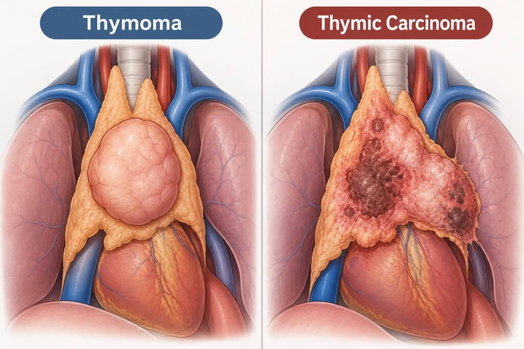
Thymoma and thymic carcinoma both begin in thymic epithelial cells but differ in behaviour and prognosis.
Thymoma is often localised at diagnosis, grows slowly and is strongly associated with myasthenia gravis. When completely removed, outcomes are often excellent.
Thymic carcinoma is more likely to invade the pericardium, great vessels or lung, and may spread to lymph nodes, liver or bone. Autoimmune syndromes are less common.
The World Health Organization classifies thymomas as types A, AB, B1, B2 and B3. Type A is generally least aggressive, while B3 carries a higher risk. Thymic carcinoma is classified separately and often requires more intensive treatment.
Causes and Risk Factors of Thymoma
The cause of thymoma is usually unknown. Unlike lung cancer, it has no established link with smoking, occupation or lifestyle.
Genetic changes in thymic epithelial cells are thought to drive tumour growth. Research continues to investigate these changes and identify possible treatment targets.
Known or suspected risk factors include:
Age: Most cases occur between ages 40 and 70, although thymoma can develop at any age.
Autoimmune background: Myasthenia gravis is particularly associated with thymoma.
Rare genetic associations: Rarely, conditions such as MEN1 or familial susceptibility may be involved.
Thymoma is not contagious and does not usually run in families.
Symptoms of Thymoma and Thymic Carcinoma
Between one-third and one-half of thymomas are discovered by chance during a chest X-ray or CT scan, and many people have no symptoms at all. When symptoms do occur, they may be caused by the tumour pressing on nearby structures. These can include a persistent cough, chest pain or a feeling of tightness behind the breastbone, breathlessness (particularly during physical activity), hoarseness, or difficulty swallowing.
Some people may also develop swelling of the face, more visible veins in the neck, or a feeling of fullness in the head if the tumour blocks the superior vena cava, a major vein that carries blood from the upper body to the heart.
If a thymoma grows into nearby tissues, it may affect the phrenic nerve, which can cause the diaphragm to become raised and lead to breathlessness. It may also affect the lining around the heart (the pericardium), causing fluid to build up around the heart.
Thymomas can also be linked to conditions affecting the immune system. One of the most common is myasthenia gravis, which can cause muscle weakness, drooping eyelids, double vision, or difficulty chewing or speaking.
Other immune-related problems can cause repeated infections or low blood cell counts. Anyone experiencing chest symptoms along with unexplained muscle weakness should seek medical assessment promptly.
"I was diagnosed in March with a rare condition. My local hospital said it was inoperable due to the size of the mass. Mr Scarci reassured me the thymoma was resectable. I was booked for surgery the following week where he successfully removed the entire tumour."
Paraneoplastic and Autoimmune Conditions Linked to Thymoma
Paraneoplastic syndromes are immune reactions caused by cancer that affect other parts of the body. Thymoma has a particularly strong association with autoimmune disease.
Key conditions include:
Myasthenia gravis (MG): Occurs in around 30 to 40 per cent of people with thymoma. Around 10 to 15 per cent of people with MG have a thymoma.
Pure red cell aplasia: The bone marrow produces too few red blood cells, causing severe anaemia.
Hypogammaglobulinaemia: Low antibody levels increase infection risk.
Other autoimmune diseases: Including thyroid disease, lupus-like syndromes and inflammatory conditions.
Thymectomy may improve myasthenia gravis, but some patients still need long-term neurological treatment. Care should involve thoracic surgery, neurology and, where needed, immunology.
How Thymoma Is Diagnosed
Diagnosis usually involves clinical assessment, imaging, blood tests and sometimes a biopsy. Findings should be reviewed by a multidisciplinary team before treatment is planned.
Imaging
A chest X-ray may identify an abnormality, but CT of the chest and upper abdomen is the main first-line test. It shows the size and position of the mass and whether it involves nearby structures.
MRI can clarify involvement of the pericardium or major blood vessels and distinguish cystic from solid masses. FDG-PET may help assess spread or distinguish thymoma from thymic carcinoma.
Blood tests
Tests usually include a full blood count and kidney and liver function. Acetylcholine receptor antibodies are important when myasthenia gravis is suspected.
Tissue diagnosis
A diagnosis may be confirmed using a CT-guided needle biopsy or mediastinoscopy. However, if imaging strongly suggests a resectable thymoma, surgery may proceed without a pre-operative biopsy in selected cases.
Staging and Classification of Thymic Tumours
Staging describes how far a tumour has spread and guides treatment and prognosis.
Masaoka–Koga Staging System
The Masaoka–Koga system remains widely used:
Stage I: The tumour is encapsulated with no invasion through its capsule.
Stage II: There is microscopic or visible invasion into surrounding fat or mediastinal pleura.
Stage III: The tumour invades nearby structures, such as the pericardium, lung or great vessels.
Stage IVa: The tumour has spread to the pleura or pericardium.
Stage IVb: There are distant metastases, including to lymph nodes, liver or bone.
TNM Staging
The AJCC TNM system is increasingly used in large centres and clinical trials. It assesses tumour extent, lymph nodes and distant spread.
WHO Histological Classification
WHO classification divides thymomas into types A, AB, B1, B2 and B3. Type A and AB tumours usually have the best outlook. B3 thymomas and thymic carcinomas have a greater risk of recurrence and may need more intensive treatment. Squamous cell carcinoma is the most common type of thymic carcinoma.
Have a question about your diagnosis?
A consultation gives you a personalised plan — not a generic estimate.
Principles of Thymoma Treatment Planning
Thymoma treatment depends on tumour type, stage, resectability, overall health and associated autoimmune disease.
The multidisciplinary team (MDT) usually includes:
Thoracic surgeon
Medical oncologist
Clinical or radiation oncologist
Radiologist
Pathologist
Neurologist, where myasthenia gravis is present
For resectable thymoma, complete surgical removal is the main treatment. Advanced or unresectable disease may require chemotherapy, radiotherapy or both.
MDTs use guidance from organisations including ESMO, NCCN and BTOG while tailoring treatment to the individual.
Early-Stage Thymoma (Stage I–II): Treatment Options
Stage I and favourable stage II thymomas are usually confined to the thymus or show limited local invasion. Complete removal offers a high chance of cure.
Surgery usually involves total thymectomy, removing the thymus and surrounding mediastinal fat. Evidence suggests this provides better long-term control than removing the tumour alone.
Postoperative radiotherapy is not recommended after complete resection of stage I thymoma. It may be considered in stage II disease where there are close or involved margins, more extensive invasion or higher-risk histology, such as B2 or B3.
VATS or robotic thymectomy may be appropriate for selected smaller, early-stage tumours. People with myasthenia gravis usually require extended thymectomy to support both cancer and neurological outcomes.
Locally Advanced Thymoma and Thymic Carcinoma (Stage III)
Stage III tumours invade nearby structures, such as the pericardium, lung or major blood vessels. Treatment often requires a combined approach.
Key points for stage III management:
Induction chemotherapy
Cisplatin-based chemotherapy may shrink the tumour before surgery and improve the chance of complete removal. Response rates are commonly around 50 to 80 per cent.
Surgery first
Selected resectable tumours may be removed first, with further treatment determined by pathology and margins.
Postoperative radiotherapy
Often considered after incomplete resection or in higher-risk stage II or III disease.
Thymic carcinoma
More often requires chemotherapy, radiotherapy and surgery because it has a higher recurrence risk.
The MDT decides the treatment sequence case by case.
Stage IV and Metastatic Thymic Tumours
Stage IVa disease has spread within the pleura or pericardium. Stage IVb disease has spread beyond the chest, such as to distant lymph nodes, liver or bone.
Treatment may include:
Systemic therapy: Cisplatin-based chemotherapy is commonly used, and thymoma can be relatively chemosensitive.
Surgery in selected cases: Pleural implants may sometimes be removed in carefully selected stage IVa cases.
Supportive and palliative care: This focuses on symptom relief, quality of life and disease control where cure is not realistic.
Clinical trials should be considered for advanced or recurrent disease.
Surgery for Thymoma and Thymic Carcinoma
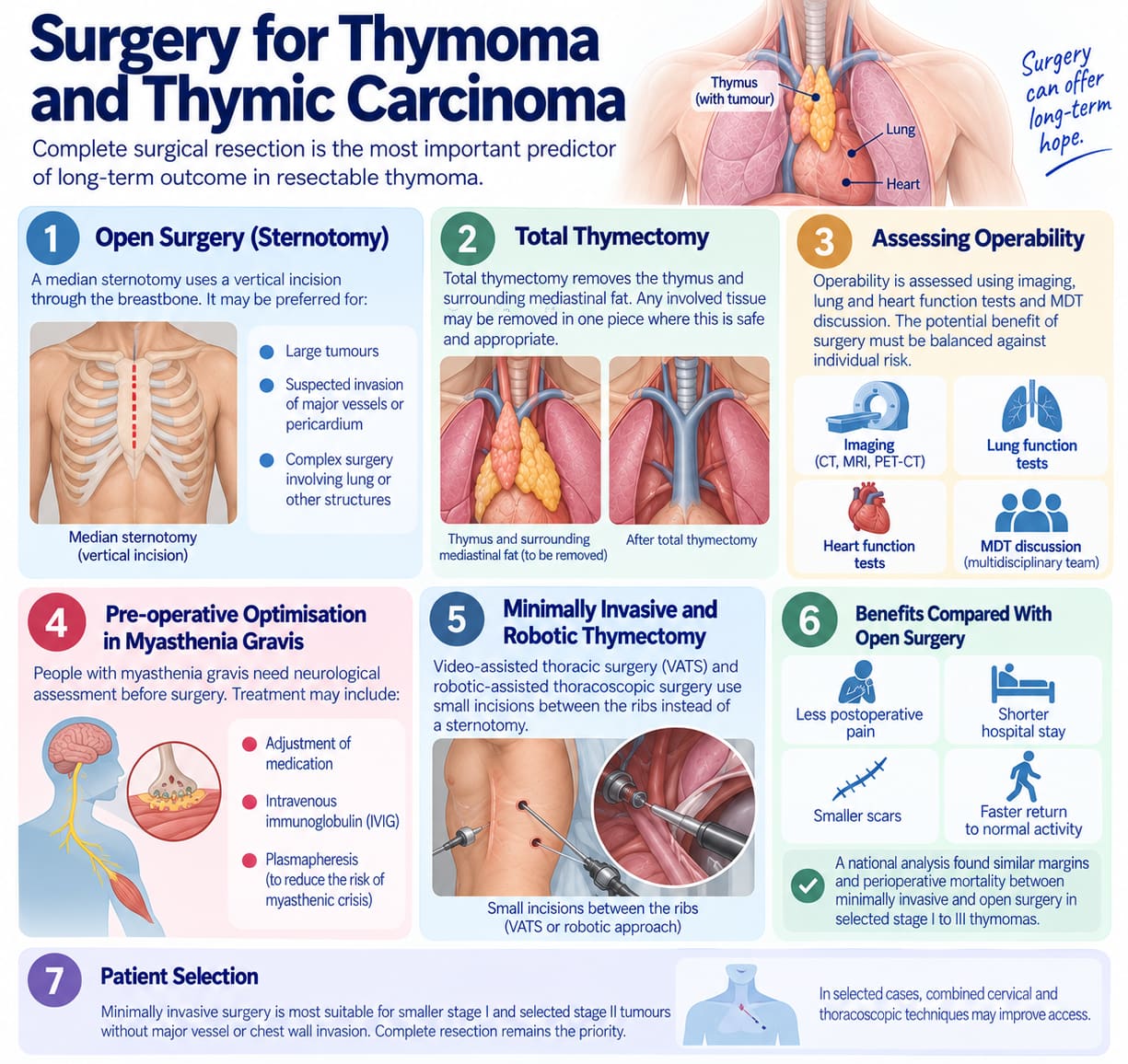
Complete surgical resection is the most important predictor of long-term outcome in resectable thymoma.
Open Surgery (Sternotomy)
A median sternotomy uses a vertical incision through the breastbone. It may be preferred for:
Large tumours
Suspected invasion of major vessels or pericardium
Complex surgery involving lung or other structures
Total Thymectomy
Total thymectomy removes the thymus and surrounding mediastinal fat. Any involved tissue may be removed in one piece where this is safe and appropriate.
Assessing Operability
Operability is assessed using imaging, lung and heart function tests and MDT discussion. The potential benefit of surgery must be balanced against individual risk.
Pre-operative Optimisation in Myasthenia Gravis
People with myasthenia gravis need neurological assessment before surgery. Treatment may include adjustment of medication, intravenous immunoglobulin or plasmapheresis to reduce the risk of myasthenic crisis.
Minimally Invasive and Robotic Thymectomy
Video-assisted thoracic surgery and robotic-assisted thoracoscopic surgery use small incisions between the ribs instead of a sternotomy.
Benefits Compared With Open Surgery
Less postoperative pain
Shorter hospital stay
Smaller scars
Faster return to normal activity
A national analysis found similar margins and perioperative mortality between minimally invasive and open surgery in selected stage I to III thymomas.
Patient Selection
Minimally invasive surgery is most suitable for smaller stage I and selected stage II tumours without major vessel or chest wall invasion. Complete resection remains the priority.
In selected cases, combined cervical and thoracoscopic techniques may improve access.
Radiation Therapy for Thymic Tumours
Radiotherapy uses high-energy radiation to destroy cancer cells. It may be given after surgery or when surgery is not possible.
When Is Radiotherapy Used?
Radiotherapy may be recommended after surgery when the tumour could not be completely removed, or for selected people with higher-risk stage II or stage III thymoma. This is known as postoperative radiotherapy and aims to reduce the chance of the cancer returning in the treated area.
For tumours that cannot be removed with surgery, radiotherapy may be given alongside chemotherapy as the main treatment approach. This is called definitive chemoradiotherapy.
Radiotherapy before surgery is used less often, but may occasionally be considered for selected locally advanced tumours. In metastatic disease, palliative radiotherapy can help relieve symptoms caused by the cancer, such as pain or pressure on nearby structures.
It is not recommended after complete resection of stage I thymoma.
Modern Techniques
Intensity-modulated radiotherapy and image-guided radiotherapy help target the tumour while reducing exposure to the heart and lungs.
Side Effects
Short-term: Fatigue, skin changes, oesophagitis and cough
Long-term: Rarely, pneumonitis, pericarditis or pulmonary fibrosis
Careful planning is especially important in younger people and those with heart or lung disease.
"Dr. Marco Scarci is a deeply knowledgeable, open-minded, and empathetic surgeon. He listens to details and solves problems to truly deliver the most optimal results. As a medical professional myself, I can tell you that finding a specialist like Dr. Scarci is very rare, and it is a privilege to have him in my corner."
Proton Beam Therapy and Advanced Radiotherapy in London
Proton beam therapy delivers radiation with limited exit dose, potentially reducing exposure to the heart, lungs and spinal cord.
When Might Proton Therapy Be Recommended?
Stage II or III disease close to critical structures
Re-irradiation for recurrent thymoma
Younger patients where long-term organ protection is particularly important
Complex thymic carcinoma
How Treatment Is Delivered
Planning usually includes CT and MRI scans, followed by daily outpatient treatment for around five to six weeks. Referral may follow assessment by a thoracic surgeon for complex chest conditions.
Proton therapy is not needed for every patient. For many, IMRT offers effective treatment with acceptable side effects.
Chemotherapy and Systemic Therapy for Thymoma and Thymic Carcinoma
Systemic therapy includes chemotherapy and selected targeted medicines that treat cancer cells throughout the body.
When Is Chemotherapy Used?
Chemotherapy may be used before surgery for locally advanced thymoma or thymic carcinoma. This is known as induction therapy and aims to shrink or control the tumour, potentially making surgery more achievable.
In selected higher-risk cases, chemotherapy may be given alongside radiotherapy or after radiotherapy. This may be considered where there is a greater risk of the cancer returning.
For tumours that cannot be removed with surgery, or where the cancer has spread to other parts of the body, chemotherapy is commonly used as the first treatment approach. The treatment plan depends on the type and stage of the tumour, previous treatments, and the person’s overall health.
Common Regimens
Cisplatin-based regimens are commonly used for thymoma. PAC combines cisplatin, doxorubicin and cyclophosphamide. Carboplatin and paclitaxel are often used for thymic carcinoma.
Side Effects
Possible side effects include nausea, hair loss, fatigue, low blood counts and a higher risk of infection. Anti-sickness medication and supportive care can reduce many of these effects.
Targeted Therapies, Immunotherapy and Emerging Treatments
Targeted therapies act on specific cancer pathways. Immunotherapy helps the immune system recognise cancer cells.
Current Evidence
EGFR expression is common in thymoma, although treatments designed to block EGFR have produced inconsistent results. Sunitinib and everolimus have shown activity in some thymic epithelial tumours, particularly thymic carcinoma.
PD-1 and PD-L1 immunotherapy medicines can produce responses in people whose disease has previously been treated. However, in thymoma, they can also cause serious immune-related side effects, so careful patient selection and close monitoring are important.
Research into new treatment combinations is continuing. The CAVEATT trial reported encouraging disease control with avelumab and axitinib in heavily pretreated B3 thymoma and thymic carcinoma.
Immunotherapy combined with chemotherapy, including approaches studied in the MARBLE trial, may also provide further options for people with advanced thymic carcinoma in the future.
Important Cautions
These treatments should be used through specialist protocols or clinical trials, with close monitoring for autoimmune complications. People with advanced or recurrent disease should ask about trial eligibility.
Not sure about your treatment options?
Mr Scarci provides expert consultations typically within one week of contact.
Corticosteroids, Octreotide and Other Adjunctive Therapies
High-dose corticosteroids can occasionally shrink thymoma and may be used as a temporary measure or when other options are unsuitable.
Octreotide, sometimes combined with prednisone, may benefit selected tumours with somatostatin receptor uptake on imaging.
These treatments are usually considered after multidisciplinary discussion within a specialist thoracic surgery team. Steroids may also help manage myasthenia gravis and immune-related side effects.
Treatment of Recurrent Thymoma and Thymic Carcinoma
Thymoma can recur years after apparently complete surgery. Reported recurrence rates vary by stage and tumour type.
Options for Localised Recurrence
Repeat surgery: Removal of isolated pleural or local recurrence may be considered in selected cases.
Targeted radiotherapy: May be used alone or with surgery.
Multimodality treatment: Some pleural recurrences require combinations of surgery, radiotherapy and systemic treatment.
A large cohort study found recurrence rates ranging from around 4 per cent in stage I to 46 per cent in stage IV disease.
Systemic Options for Recurrent Disease
Further platinum-based chemotherapy
Medicines such as pemetrexed, paclitaxel or docetaxel
Clinical trials of targeted or immunotherapy combinations
Treatment should be individualised according to previous treatment, recurrence site, symptoms and overall health.
Thymoma, Myasthenia Gravis and Anaesthetic Planning
Myasthenia gravis requires particular care before, during and after surgery.
Pre-operative Optimisation
Before surgery, patients with myasthenia gravis will usually have a review with their neurology team to ensure their condition is as stable as possible. This may include adjusting anticholinesterase medicines and reviewing steroid or immunosuppressant treatment.
For some patients, particularly where symptoms are not well controlled or surgery is expected to carry a higher risk, intravenous immunoglobulin (IVIG) or plasmapheresis may be considered before the operation. The anaesthetic and surgical teams will use this information to plan care carefully and reduce the risk of complications around surgery.
Anaesthetic Considerations
People with myasthenia gravis can be highly sensitive to certain muscle relaxants. Some require high-dependency or intensive care monitoring after major surgery.
Experienced teams plan anaesthesia, pain management and postoperative respiratory support carefully to reduce the risk of myasthenic crisis.
Postoperative Recovery and Short-Term Complications
Recovery after thymoma surgery depends on the operation performed.
VATS or Robotic Thymectomy
Hospital stay is often two to four days
Earlier mobilisation and return to daily activity
Less pain and faster wound healing
Open Sternotomy
Hospital stay is often five to ten days
More discomfort around the breastbone
Longer recovery before unrestricted activity
Common Short-Term Issues
Pain
Temporary breathlessness
Fatigue
Wound care needs
Possible Complications
Bleeding
Infection
Phrenic nerve injury
Rare injury to major blood vessels
Phrenic nerve injury may occasionally require diaphragmatic plication. Physiotherapy, good pain control and smoking cessation can reduce respiratory complications.
Long-Term Follow-Up and Monitoring for Recurrence
Thymoma can recur 10 to 20 years after treatment. Long-term surveillance is therefore important.
Typical Follow-Up Schedule
CT of the chest every 6 to 12 months for the first few years
Annual CT thereafter, tailored to stage, pathology and previous treatment
Regular review for symptoms or autoimmune complications
PET-CT may be used when CT findings are uncertain or distant spread is suspected.
Prognosis and Survival Outlook
Prognosis for thymoma depends primarily on three factors: stage at diagnosis, completeness of surgical resection, and histological type.
Approximate Five-Year Survival by Stage
Stage | Five-Year Survival |
Stage I (completely resected) | Approximately 90 per cent. Five-year survival for completely resected stage I thymoma exceeds 90 per cent. |
Stage II | Approximately 70 to 90 per cent |
Stage III | Approximately 50 to 70 per cent |
Stage IV | Approximately 30 to 50 per cent |
These figures are broad benchmarks, not an individual prediction. Type A and AB thymomas generally have lower recurrence risk than B3 thymoma and thymic carcinoma. Many people with early-stage disease are cured, while some people with advanced disease achieve long-term control with combined treatment.
Living With and Beyond Thymoma
A rare cancer diagnosis can feel isolating. Clear information, emotional support and contact with others affected by thymic tumours can help.
Strategies for Coping
It can help to ask your healthcare team for clear information about your diagnosis, treatment and what to expect at each stage. If it feels right, consider bringing a family member or friend to appointments so they can provide support and help remember important details.
Counselling or psychological support may also help, particularly if you feel anxious, low, or overwhelmed. Connecting with people who have had similar experiences can be reassuring too. Support groups such as ThymicUK may offer information and a sense of community.
Lifestyle and Recovery
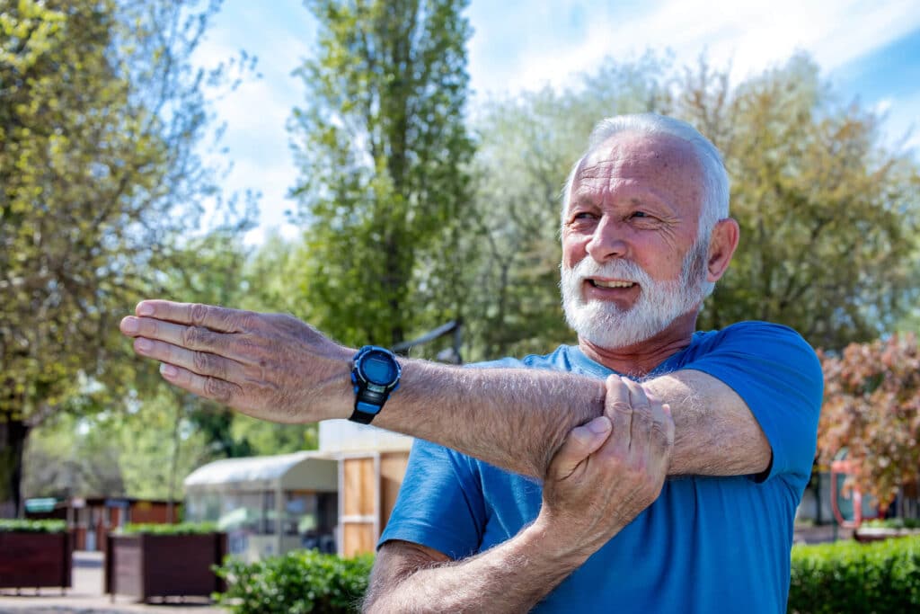
Recovery looks different for everyone. Stopping smoking, eating a balanced diet and gradually building activity through gentle exercise or physiotherapy can all support general health and recovery. Your clinical team can advise on a suitable pace, especially after surgery or during ongoing treatment.
If your immune system is weakened, ask your healthcare team for personalised vaccination advice. People taking long-term steroid medication may also need to discuss bone health and whether monitoring or preventative support is appropriate.
Many people return to work and everyday activities after treatment, although the pace and experience of recovery vary from person to person. Patient experiences can offer helpful perspective, but everyone’s journey with thymoma is different.
Why Choose Mr Marco Scarci for Thymoma and Thymic Carcinoma Care in London
Thymoma should be managed by a team experienced in rare mediastinal tumours, minimally invasive surgery, and myasthenia gravis.
Mr Marco Scarci is a consultant thoracic surgeon in London who provides assessment, surgical treatment and second opinions for thymoma and thymic carcinoma. His practice offers access to multidisciplinary care, VATS and robotic approaches where appropriate, and review of complex or borderline-operable cases.
For an individual assessment or second opinion, contact Mr Scarci’s clinic to arrange a thoracic surgery consultation.
Frequently Asked Questions About Thymoma Treatment
Is thymoma the same as lung cancer?
No. Thymoma develops in the thymus, not the lung. It has different risk factors, behaviour and treatment. CT, PET-CT and sometimes biopsy help distinguish thymic tumours from lung cancer and other chest masses.
Will removing my thymus gland affect my immune system?
Usually not significantly in adults. The thymus has largely completed its immune-development role by adulthood. Infection risk after treatment is more often related to chemotherapy, steroids or an associated immune condition.
Do all thymoma patients need chemotherapy or radiotherapy after surgery?
No. Many people with completely resected stage I thymoma need no treatment beyond surgery. Further treatment may be considered for higher-stage disease, thymic carcinoma, positive margins or other high-risk features.
Can thymoma come back after successful treatment?
Yes. Recurrence may occur years later, which is why long-term imaging follow-up is needed. Repeat surgery, radiotherapy or systemic treatment may be possible depending on the pattern of recurrence.
How quickly can I get a specialist opinion if I have just been diagnosed?
Urgency depends on symptoms, imaging findings and whether there is concern about compression of major structures or myasthenia gravis. Bring all available CT, MRI, PET-CT images and reports to a specialist appointment so the treatment plan can be reviewed efficiently.
