

A chest wall deformity can affect how the chest looks, how comfortably someone breathes and exercises, and how they feel about their body. For some people, the difference in chest shape is subtle and causes few problems. For others, it can produce physical limitations, discomfort or significant self-consciousness.
Chest wall abnormalities range from common conditions such as pectus excavatum and pectus carinatum to rarer syndromic and acquired deformities. Treatment depends on the type of deformity, its severity, the patient’s age, symptoms and whether the heart, lungs or daily life are affected.
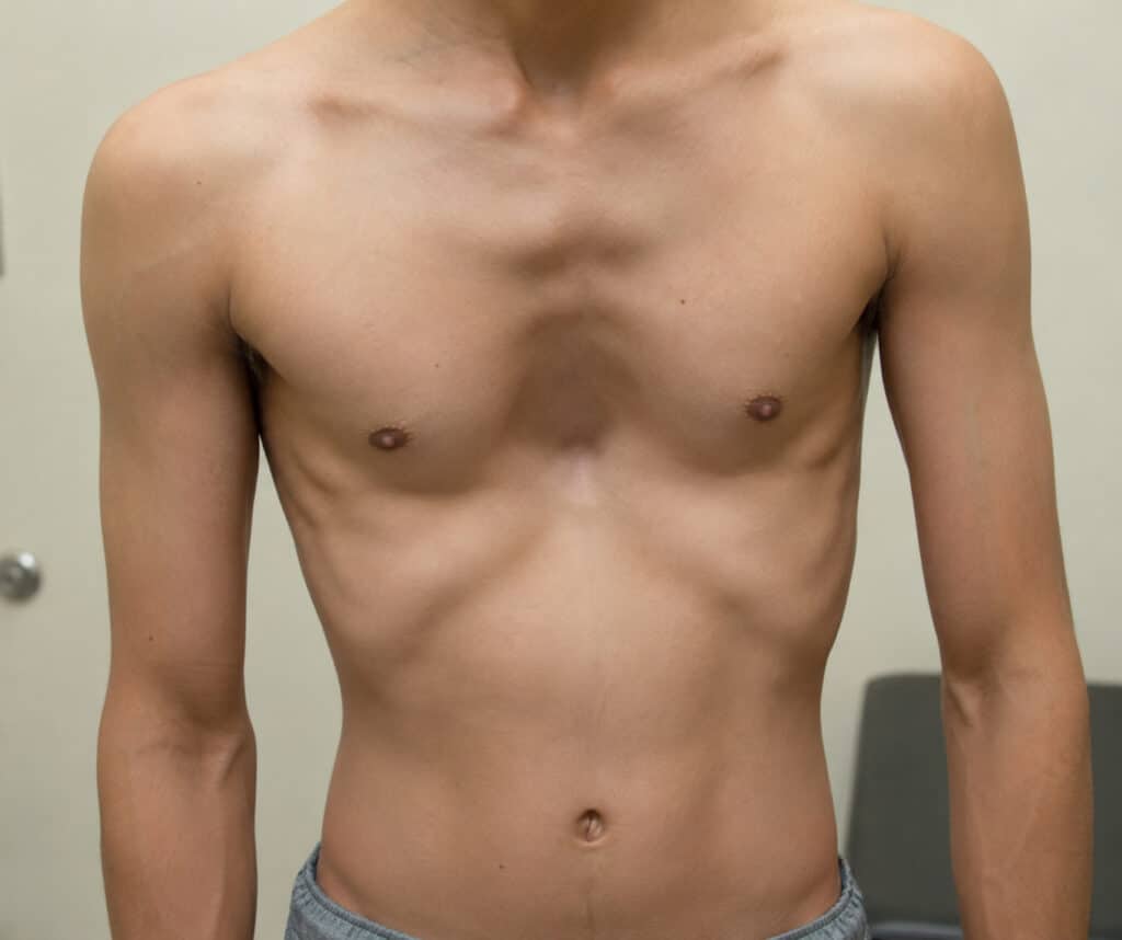
A chest wall deformity is an abnormal shape of the sternum, ribs or costal cartilage. Most are congenital and often become more noticeable during childhood growth spurts and puberty.
Pectus excavatum (funnel chest) and pectus carinatum (pigeon chest) are the two most common deformities of the anterior chest wall, with pectus excavatum accounting for the majority of cases.
Chest wall deformities can range from cosmetic variations to significant health problems. Breathlessness, chest pain and reduced exercise tolerance may occur, while effects on confidence and body image can also be substantial.
Treatment ranges from observation and targeted exercises to bracing, vacuum bell therapy and surgery, depending on deformity type, severity, age and symptoms.
What Is a Chest Wall Deformity?
A chest wall deformity is an abnormal shape or development of the structures forming the front, sides or overall rib cage. These include the sternum, ribs, costal cartilage connecting the ribs to the breastbone and, in some conditions, the spine.
During normal development, these structures grow together to form a protective cage around the heart and lungs. When chest wall development is altered, the result may range from barely noticeable asymmetry to a significant structural abnormality.
Most chest wall deformities in otherwise healthy patients are congenital, meaning they are present from birth even if they are not recognised until later. Others develop after trauma or surgery, chronic lung disease or neuromuscular conditions that gradually affect posture and chest shape.
Common visible patterns include:
Sunken chest, usually pectus excavatum or funnel chest
Protruding chest, usually pectus carinatum or pigeon chest
Mixed patterns combining depression and protrusion
Marked asymmetry or underdeveloped chest muscles, as in Poland syndrome
Many patients first notice these changes during childhood or puberty, when rapid skeletal growth can make an existing difference in chest shape much more obvious.
Not sure about your treatment options?
Mr Scarci provides expert consultations typically within one week of contact.
How Common Are Chest Wall Deformities?
Chest wall deformities are more common than many people realise. Mild cases may never be formally diagnosed because they are attributed to posture or cause few symptoms.
Overall, they are estimated to occur in approximately 1 in 300 to 400 live births. Pectus excavatum accounts for roughly 90% of common anterior chest wall deformities and affects about 1 in 300 to 400 people.
Pectus carinatum is approximately 2 to 4 times less common than pectus excavatum, with an estimated incidence of around 1 in 1,500. Both conditions show a male predominance, with reported ratios ranging from approximately 3:1 to 5:1.
Much rarer conditions include Poland syndrome, estimated at around 1 in 20,000 to 30,000 live births, and sternal clefts, which account for only about 0.15% of chest wall deformities. Ectopia cordis is exceptionally rare, with estimates ranging from approximately 1 in 5.5 to 7.9 million births.
What Causes Chest Wall Deformities?
The precise origins of congenital chest wall abnormalities are not fully understood. Most chest deformities are thought to involve altered development of the costal cartilages that connect the ribs to the sternum, resulting in either inward depression, as in pectus excavatum, or outward protrusion, as in pectus carinatum. However, the underlying process appears to be more complex than simple cartilage overgrowth.
Biological Mechanisms
Abnormal cartilage development is considered an important mechanism behind many pectus conditions. Traditionally, the prevailing theory has been that the costal cartilages grow disproportionately relative to the surrounding thoracic cage, pushing the sternum inward or outward.
However, more recent three-dimensional CT studies have challenged this simple overgrowth model. In some patients with pectus excavatum, the cartilage is not necessarily longer than normal and may even be shorter. This suggests that cartilage quality, stiffness and collagen composition may be as important as cartilage length.
Key biological factors include:
Connective tissue and collagen abnormalities: patients with pectus excavatum may show disorganised collagen fibre arrangement, altered proteoglycan content, mineral differences and reduced biomechanical strength of the cartilage. Weakened cartilage may be less able to resist normal respiratory forces and diaphragm traction, contributing to gradual deformation.
Growth plate disturbances: abnormal curvature, angulation or torsion of the sternum, together with changes in sternal growth plates during childhood, can alter how the sternum responds to forces from the ribs and diaphragm.
Mechanical forces during growth spurts: deformities often become much more noticeable during preadolescence and puberty, when the chest wall is exposed to rapid skeletal growth, respiratory forces and changes in posture.
Genetic Influences
Genetic factors are also associated with chest wall deformities. Roughly 40% to 50% of pectus excavatum cases show some degree of familial clustering, with affected relatives sometimes appearing across several generations.
Reported inheritance patterns include autosomal dominant, autosomal recessive and X-linked patterns, although many families do not follow a clear Mendelian pattern.
Several genes and biological pathways have been investigated, including:
GPR126, linked to pectus excavatum and scoliosis in experimental models
COL5A1, COL1A1 and COL27A1, which influence collagen and connective tissue structure
SMAD4 and TGFB3, involved in TGF-β/BMP signalling and skeletal development
GAL3ST4 and TINAG, which may influence extracellular matrix composition and cell adhesion
Chest wall deformities are also associated with recognised connective tissue and genetic conditions, including Marfan syndrome, Ehlers-Danlos syndrome and Noonan syndrome. In these patients, the chest deformity may form part of a broader pattern of skeletal and soft-tissue differences.
These associations are also relevant when assessing pectus excavatum causes, particularly where there is a strong family history or other connective tissue features.
Embryological and Syndromic Causes
Some syndromic chest wall deformities develop because of disturbances during early embryological development.
Poland syndrome, for example, is thought to result from disruption of blood flow through the subclavian artery during approximately the sixth to seventh week of gestation. This can lead to underdevelopment of chest muscles, ribs and surrounding soft tissues.
Jeune syndrome and Jarcho-Levin syndrome involve skeletal development abnormalities that affect the ribs, spine, and overall thoracic shape. These conditions are congenital and are not caused by anything parents did or did not do during pregnancy.
Acquired Causes
Not all chest wall deformities are congenital. Structural changes can also develop later in life because of injury, surgery or underlying disease.
Acquired causes include previous thoracic or cardiac surgery, including open-heart surgery during childhood; rib fractures or severe blunt chest trauma, which may require specialist rib injury treatment if pain or deformity persists; severe chronic lung disease leading to barrel chest; neuromuscular disorders that gradually alter posture and chest wall mechanics; and tumours within the mediastinum or chest wall that distort the thoracic cavity from within.
Want a specialist opinion on your condition?
Get a clear answer from a surgeon who will be with you throughout.
Types of Chest Wall Deformities
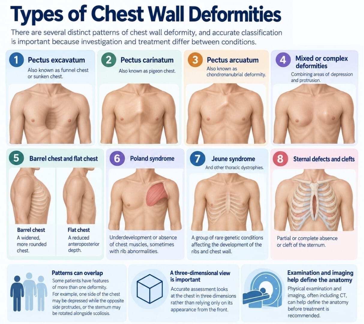
There are several distinct patterns of chest wall deformity, and accurate classification is important because investigation and treatment differ between conditions.
The main categories include:
Pectus excavatum, also known as funnel chest or sunken chest
Pectus carinatum, also known as pigeon chest
Pectus arcuatum, or chondromanubrial deformity
Mixed or complex deformities, combining areas of depression and protrusion
Barrel chest and flat chest
Poland syndrome
Jeune syndrome and other thoracic dystrophies
Sternal defects and clefts
Some patients have features of more than one pattern. For example, one side of the chest may be depressed while the opposite side protrudes, or the sternum may be rotated alongside scoliosis.
Accurate assessment therefore looks at the chest in three dimensions rather than relying only on its appearance from the front. Physical examination and imaging, often including CT, can help define the anatomy before treatment is recommended.
Pectus Excavatum: Sunken or Funnel Chest
Pectus excavatum causes central or asymmetric depression of the sternum and adjacent costal cartilage, creating the characteristic sunken or funnel-shaped appearance.
It is the most common congenital anterior chest wall deformity and becomes particularly noticeable during puberty. Common symptoms include shortness of breath on exertion, chest tightness, palpitations, exercise intolerance, fatigue and chest or back discomfort.
In more severe cases, the inward position of the sternum can reduce the available space for the heart and lungs. CT imaging may be used to calculate the Haller Index, with a value of approximately 3.25 or higher commonly associated with more severe pectus excavatum.
Associated conditions can include scoliosis, mitral valve prolapse, aortic root dilatation and connective tissue disorders.
Pectus Carinatum: Pigeon Chest
Pectus carinatum is an outward protrusion of the sternum and costal cartilage, producing the characteristic pigeon-chest appearance. It is the second most common chest wall deformity and occurs approximately 2 to 4 times less frequently than pectus excavatum.
The deformity often becomes obvious during later childhood or early adolescence. Unlike severe pectus excavatum, pectus carinatum usually causes relatively little direct heart or lung impairment, although some patients experience mild breathlessness or chest discomfort during sport.
Its psychological effects can be considerable. Prominence of the sternum may cause self-consciousness, avoidance of swimming or changing rooms and difficulty wearing fitted clothing.
In children and adolescents with flexible chest walls, pectus carinatum bracing is often the preferred first-line treatment. Controlled external pressure gradually reshapes the chest over time, while surgery is generally reserved for rigid deformities, adults, or patients in whom bracing is unsuccessful.
Pectus Arcuatum and Mixed Deformities
Pectus arcuatum, also called a chondromanubrial deformity, is a less common pattern in which the upper sternum protrudes while the lower sternum may remain flat or slightly depressed.
Because its appearance can resemble both pectus excavatum and pectus carinatum, accurate diagnosis is important. Treating it as a standard pectus deformity without recognising the underlying anatomy can produce suboptimal results.
Mixed deformities may combine depression, protrusion and rotational asymmetry across different parts of the chest. These cases often require detailed three-dimensional planning and sometimes a combination of open and minimally invasive techniques.
Barrel Chest and Flat Chest
Not every abnormal chest shape falls within the pectus group.
Barrel chest describes an increased front-to-back diameter of the thorax, giving the chest a rounded appearance. It is commonly associated with advanced emphysema and chronic obstructive pulmonary disease, where long-term air trapping gradually alters chest mechanics.
Flat chest describes a reduced front-to-back diameter relative to chest width. It may occur on its own or alongside pectus excavatum.
Because these patterns may result from underlying respiratory disease rather than isolated cartilage abnormalities, treatment often focuses on improving lung health, posture and breathing mechanics rather than surgically changing the chest wall.
Poland Syndrome and Rare Syndromic Deformities
Poland syndrome is a rare congenital condition characterised by partial or complete absence of the pectoralis major muscle on one side of the chest. Rib abnormalities and hand differences, such as shortened fingers, may also occur.
The condition is estimated to affect approximately 1 in 20,000 to 30,000 births and appears more frequently on the right side. Chest asymmetry may be relatively subtle during childhood but can become more obvious during puberty.
Treatment is highly individualised and may involve reconstructive surgery after growth is largely complete, using muscle flaps, implants or autologous fat grafting depending on the anatomy and goals of the patient.
Other rare syndromic chest wall abnormalities include:
Condition | Key Features |
Jeune syndrome | Very narrow, rigid chest with short ribs and potentially severe respiratory compromise |
Jarcho-Levin syndrome | Multiple vertebral and rib abnormalities producing a shortened, abnormal chest shape |
Sternal clefts | Failure of normal sternal fusion, sometimes with visible underlying cardiac pulsation |
Cantrell’s pentalogy | Combination of midline sternal, diaphragmatic and cardiac abnormalities |
Ectopia cordis | Extremely rare condition in which the heart lies partly or completely outside the normal chest cavity |
These conditions are uncommon but can cause significant restriction of chest volume and lung development. Severe cases require assessment in highly specialised multidisciplinary centres.
Have a question about your diagnosis?
A consultation gives you a personalised plan — not a generic estimate.
Jeune Syndrome and Thoracic Dystrophies
Jeune syndrome, or asphyxiating thoracic dystrophy, is a rare autosomal recessive condition characterised by a very narrow, rigid chest and short ribs. It occurs in approximately 1 in 120,000 births.
The restricted chest cavity limits lung expansion and can cause significant breathing difficulties during infancy and childhood. Respiratory infections can therefore become particularly serious.
Selected children may undergo thoracic expansion procedures or implantation of expandable devices designed to increase the available space for lung growth.
Jarcho-Levin syndrome similarly involves multiple vertebral and rib abnormalities and can produce severe respiratory compromise. These conditions require coordinated care involving paediatric, respiratory, genetic and surgical specialists.
Typical Symptoms and When to Worry
Many people with mild chest wall deformities have few or no physical symptoms. As severity increases, however, the abnormal chest shape can begin to affect breathing, exercise and musculoskeletal comfort.
Common symptoms include breathlessness on exertion, chest tightness, palpitations, fatigue, reduced exercise capacity, chest or back pain and occasional fainting during intense activity.
More concerning changes include rapidly worsening deformity during a growth spurt, new limitations in sports performance, exertional chest pain, recurrent respiratory infections, or visible abnormal movement or pulsation beneath the chest wall.
Persistent chest wall pain should also be assessed when it is worsening, unexplained or associated with a structural abnormality.
Children with very narrow thoracic cages or major sternal defects can present much earlier with rapid breathing, poor growth or respiratory compromise and require prompt specialist review.
Psychological and Social Impact
The psychological effect of a visible chest wall difference can be as important as the physical symptoms, particularly during adolescence and early adulthood.
Some patients avoid changing rooms, swimming pools, fitted clothing or intimate situations. Others withdraw from sport and social activities because they fear being stared at or judged.
Studies using pectus-specific quality-of-life questionnaires have found meaningful improvements following successful bracing or surgical correction in appropriately selected patients.
Psychological distress should therefore be discussed openly rather than dismissed as purely cosmetic. Where anxiety, low mood or body-image concerns become significant, counselling or psychological support may be appropriate alongside physical treatment.
How Are Chest Wall Deformities Diagnosed?
Diagnosis starts with a detailed clinical assessment. The clinician considers when the deformity first appeared, whether it is progressing, family history, related medical conditions and the effect on daily activities and emotional wellbeing.
Physical examination assesses chest wall symmetry, sternum position, ribs, posture and spinal alignment. Features suggesting scoliosis or a connective tissue disorder may prompt further investigation.
Imaging can include chest X-ray, CT scanning and MRI. CT is particularly useful for defining three-dimensional anatomy and calculating measurements such as the Haller Index in pectus excavatum.
Functional testing may include spirometry, lung-volume measurements, echocardiography and cardiopulmonary exercise testing where symptoms suggest impaired heart or lung performance.
Genetic assessment may be appropriate when the chest wall abnormality occurs alongside tall stature, unusual joint flexibility, limb differences or other syndromic features.
Non-Surgical Treatment Options
Many patients, particularly children and teenagers with flexible chest walls, can be managed without surgery.
Approach | Best Suited For | Details |
Observation | Mild deformities in growing children | Regular review to monitor progression |
Posture and physiotherapy | Mild deformities and supportive treatment | Improves chest expansion, strength and spinal alignment |
Vacuum bell therapy | Selected pectus excavatum patients | Uses suction to gradually elevate the sternum |
External bracing | Flexible pectus carinatum | Applies controlled pressure to reshape the chest |
Vacuum bell therapy works best in younger patients with flexible chest walls and usually requires regular use over months or years. Outcomes depend heavily on deformity severity and adherence.
Pectus carinatum bracing similarly requires consistent wear and regular adjustment. Structural correction is more difficult once the chest wall becomes rigid in adulthood.
Even where non-surgical treatment cannot fully alter the underlying chest shape, physiotherapy and posture work can still improve breathing mechanics, muscle strength and overall appearance.
Surgical Treatment Options
Surgery is considered when there is significant functional impairment, marked structural deformity, severe psychological impact or failure of appropriate conservative treatment.
The main categories include minimally invasive bar procedures, modified Ravitch operations and complex chest wall reconstruction.
The Nuss procedure is widely used for pectus excavatum, while selected pectus carinatum patients may undergo minimally invasive anterior bar techniques. More complicated or rigid deformities may require open reconstruction.
Modern thoracic surgery increasingly uses thoracoscopic guidance, improved implant systems, advanced imaging and enhanced recovery pathways. These changes are part of wider advances in thoracic surgery that have allowed more chest wall procedures to be planned and performed with greater precision.
The Nuss Procedure for Pectus Excavatum
The Nuss procedure is a minimally invasive operation for correcting pectus excavatum. It became widely adopted after its introduction in the late 1990s.
Two small incisions, usually around 2 to 3 cm, are made on either side of the chest. A thoracoscope allows the surgical team to visualise the inside of the chest while a curved bar is passed behind the sternum.
The bar is then rotated, pushing the sternum forward into a corrected position, and stabilisers are used to help prevent displacement.
No costal cartilage is routinely removed, and the operation avoids the larger central incision associated with traditional open repair.
Hospital stay is typically around 3 to 5 days, with pain management, breathing exercises and early mobilisation forming an important part of recovery. The bar remains in place for approximately 2 to 3 years before removal.
Different pectus excavatum treatments may be appropriate depending on chest-wall flexibility, severity and symptoms, so the Nuss procedure is not automatically the right option for every patient.
"Dr. Marco Scarci is a deeply knowledgeable, open-minded, and empathetic surgeon. He listens to details and solves problems to truly deliver the most optimal results. As a medical professional myself, I can tell you that finding a specialist like Dr. Scarci is very rare, and it is a privilege to have him in my corner."
Open and Hybrid Procedures
The modified Ravitch procedure is an open operation that involves reshaping or removing abnormal sections of costal cartilage and repositioning the sternum.
Modern modifications aim to preserve as much normal cartilage as possible while providing stable correction. Open or hybrid approaches may be preferable for very rigid adult chest walls, severe asymmetry, pectus arcuatum, recurrence after previous surgery or complex anatomical defects requiring direct reconstruction.
Recovery is generally longer than after a straightforward minimally invasive procedure because more extensive chest wall dissection is required.
Thoracic surgeons choose among minimally invasive, open, and hybrid approaches based on individual anatomy rather than using a single technique for every patient. Complex cases may also involve a multidisciplinary thoracic team.
Surgical Options for Pectus Carinatum
Bracing remains the preferred treatment for many children and teenagers with flexible pectus carinatum. Surgery is usually considered when the deformity is rigid, conservative treatment fails, or the patient presents later in adulthood.
Options include a modified Ravitch procedure, which reshapes the sternum and costal cartilage, and minimally invasive anterior bar techniques in which a bar applies pressure from the front of the chest.
The primary benefits are usually improved chest contour, symmetry and confidence, as many patients with pectus carinatum already have relatively normal heart and lung function.
Complex Chest Wall Reconstruction
Some patients require bespoke reconstruction because of severe mixed deformities, previous failed operations, major trauma, tumour resection or syndromic conditions.
Techniques can include titanium plates, mesh, custom three-dimensional implants, muscle flaps and autologous fat grafting.
Pre-operative CT reconstruction and virtual surgical planning can help surgeons design implants around the patient’s individual anatomy.
These procedures are more extensive than standard pectus surgery and are usually performed within specialist thoracic units. In these cases, the goal may include restoring chest stability and protecting underlying organs as well as improving appearance.
Risks, Complications and Long-Term Outcomes
Every surgical procedure carries risks, and these need to be discussed honestly before treatment.
Potential complications of chest wall surgery include:
Bleeding or wound infection
Pneumothorax
Bar displacement or migration
Rare metal sensitivity
Very rarely, injury to the heart, lung or major blood vessels
Modern thoracoscopic guidance, improved stabilisation techniques and careful surgical planning have reduced serious complications.
Recurrence can occur if correction is performed too early, an implant is removed prematurely, or an underlying connective tissue disorder continues to affect the chest wall.
Published series report pectus excavatum surgical success rates of approximately 86% to 98%. Most long-term studies report high patient satisfaction, sustained cosmetic improvement and, in selected symptomatic patients, better exercise tolerance and quality of life.
Life After Treatment: Recovery, Exercise and Lifestyle

Recovery varies by procedure, age, and extent of reconstruction. After uncomplicated minimally invasive pectus surgery, the early weeks focus on pain control, breathing exercises and gradual mobilisation.
During weeks 1 to 3, walking and gentle movement are encouraged. Between approximately weeks 3 and 6, many patients begin returning to school, university or desk-based work.
Progressive aerobic exercise can often resume over months 2 to 3, while return to contact sport is delayed until the chest wall has healed adequately and the bar is stable.
Avoid heavy lifting, excessive twisting, and lying directly on the front of the chest during early recovery.
Patients generally adapt well to living with a pectus bar. Normal travel and airport security are usually possible, although carrying documentation confirming the implant can be useful.
Once the bar is removed, usually after 2 to 3 years, most patients return fully to unrestricted activity.
Related Chest Wall Problems
Chest wall abnormalities do not always fit neatly into one diagnosis. Rib flaring, for example, may occur alongside pectus excavatum or pectus carinatum and can affect posture, breathing mechanics and appearance.
Some patients also present with persistent rib pain, previous chest trauma or other chest wall abnormalities that require assessment alongside the visible deformity.
Chest Wall Deformity Care With Mr Marco Scarci
Mr Marco Scarci is a consultant thoracic surgeon based in London with experience treating pectus excavatum, pectus carinatum, complex chest wall deformities, rib conditions and reconstructive thoracic problems. His current clinical scope includes minimally invasive Nuss correction, carinatum and arcuatum surgery, and complex reconstruction using custom implants where appropriate.
Assessment may include an examination of chest shape and posture, review of CT or MRI imaging, lung function testing, and cardiac investigations, depending on symptoms.
Treatment planning considers physical function, anatomy, cosmetic goals and psychological wellbeing rather than focusing on appearance or imaging measurements alone.
Private patients can arrange assessment through Mr Scarci’s London clinic. Related chest wall concerns can also be evaluated alongside the primary deformity when necessary.
Frequently Asked Questions
Can a mild chest wall deformity get worse over time?
Many mild deformities become more noticeable during growth spurts in early adolescence as the sternum and ribs lengthen and cartilage growth accelerates. After skeletal maturity, typically in the late teens to early twenties, chest shape is less likely to change dramatically, although posture, muscle tone and body composition can still influence appearance. Periodic review during childhood and adolescence can help document progression and guide the timing of treatment.
Is a chest wall deformity just a cosmetic problem?
No. Appearance is an important concern for many patients, but severe pectus excavatum and some thoracic dystrophies can affect heart or lung function. Symptoms may include shortness of breath, exercise intolerance, palpitations and chest pain. Psychological distress and low self-esteem are also legitimate health concerns and should be considered alongside physical symptoms.
What age is best for surgery to correct pectus excavatum or pigeon chest?
Many centres perform pectus surgery during the teenage years, often around ages 12 to 16, when the chest wall remains flexible but much of skeletal growth has already occurred. Adults can also benefit from surgery, although the technique and recovery may differ because the chest wall becomes more rigid with age. Timing should therefore be individualised.
Will I always need surgery, or can exercises fix my chest shape?
Targeted exercise and physiotherapy can improve posture, muscle definition, and breathing mechanics but usually do not change the underlying bone and cartilage structure in moderate to severe deformities. In milder cases, posture work, vacuum bell therapy or bracing may provide satisfactory improvement. Treatment depends on deformity type, flexibility, symptoms and individual goals.
Is it safe to travel and go through airport security with a pectus bar in place?
Most patients with pectus bars travel normally. Cabin pressure does not affect the bar or chest wall. Stainless steel or titanium implants may occasionally trigger airport security systems, so carrying documentation from the surgical team confirming the presence of the implant can be helpful.
