

Many conditions mistaken for lung cancer cause similar symptoms or scan findings. A persistent cough, chest pain, shortness of breath and weight loss can result from infection, chronic lung disease, heart disease or inflammation. An accurate diagnosis requires the full clinical picture, not one symptom or scan.
Lung cancer symptoms such as persistent cough, chest pain, shortness of breath and unexplained weight loss have many possible causes.
Common conditions mistaken for lung cancer include pneumonia, chronic obstructive pulmonary disease, asthma, benign lung nodules, pulmonary embolism and inflammatory lung disease.
A chest X-ray alone may not provide a correct diagnosis. CT scans, positron emission tomography and tissue sampling may be needed.
Lung cancer misdiagnosis can delay appropriate treatment, while a suspected cancer diagnosis can lead to unnecessary procedures when a lesion is benign.
Why Lung Cancer Is So Often Confused With Other Conditions
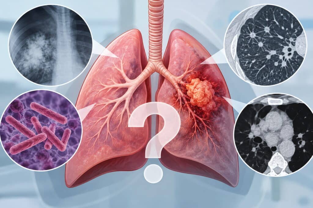
Lung cancer is a leading cause of cancer death worldwide, but its early signs are often mild and non-specific. Symptoms may develop gradually and be mistaken for a chest infection, chronic bronchitis or another lung condition.
Key overlapping lung cancer symptoms include:
A persistent cough lasting more than three weeks
Coughing up blood
Chest pain, particularly on deep breathing
Shortness of breath or difficulty breathing
Frequent lung infections
Unexplained weight loss and fatigue
Infections, lung inflammation, heart failure and obstructive lung diseases can produce similar symptoms. On imaging tests, nodules, scarring, fluid and collapse of part of the lung may resemble cancer. Doctors therefore consider medical history, including cigarette smoking and asbestos exposure, alongside examination, imaging and tissue sampling.
Pneumonia and Recurrent Chest Infections
Pneumonia is a bacterial, viral, or fungal infection that affects the air sacs. It can cause cough, sputum, chest pain, shortness of breath and a mild fever.
Pneumonia symptoms often overlap with lung cancer symptoms. Most infections improve after treatment, but persistent or recurrent pneumonia in the same area should be investigated, especially in an older person with a smoking history. An obstructing tumour can cause repeated infection behind it.
On a chest X-ray or CT scan, consolidation, collapse or a rounded shadow can resemble a mass. If an apparent pneumonia does not clear on follow-up imaging, further investigation may lead to a cancer diagnosis.
Chronic Obstructive Pulmonary Disease (COPD) and Other Obstructive Lung Diseases
Chronic obstructive pulmonary disease includes chronic bronchitis and emphysema. It is a chronic lung disease commonly linked to cigarette smoking and occupational exposures, affecting the bronchial tubes and lung tissue.
Symptoms may include chronic cough, sputum, wheezing, chest tightness, fatigue, weight loss and shortness of breath on exertion. Bronchitis and lung cancer can also coexist, so a COPD diagnosis does not rule out developing lung cancer.
People with COPD have a higher risk of lung cancer, including those developing COPD without a smoking history. One study reported that around 1% of COPD patients receive a lung cancer diagnosis each year.
CT scans may show scarring, bullae or cystic changes that can obscure lung nodules. A change in a usual cough, worsening breathing difficulties, new chest pain or symptoms that no longer respond to inhalers should prompt review.
Asthma, Allergies and Other Inflammatory Airway Problems
Asthma is a disease caused by inflammation and narrowing of the bronchial tubes in the respiratory system. Unlike lung cancer, asthma usually improves with inhalers and corticosteroids.
Both conditions can cause wheeze, chest tightness, cough, trouble breathing and recurrent chest infection. New asthma-like symptoms in adulthood, particularly in a smoker, should not automatically be assumed to be asthma without appropriate imaging.
A good response to inhalers and normal imaging favour asthma. However, coughing up blood, unexplained weight loss, or persistent symptoms despite treatment require further assessment.
Benign Lung Nodules, Scarring and Cystic Lung Diseases
Lung nodules are small spots found on CT scans. Most are benign and cause no symptoms. Common causes include:
Healed infections, including granulomas
Hamartomas
Scarring after pneumonia or tuberculosis
Inflammatory lesions
Cystic lung diseases, including emphysematous bullae, lymphangioleiomyomatosis, pulmonary Langerhans cell histiocytosis and Birt-Hogg-Dubé syndrome, can cause cysts, chest discomfort or a collapsed lung.
Radiologists assess a nodule’s size, growth, edges, density and location. Midway through the diagnostic pathway, Mr Marco Scarci, a London-based consultant thoracic surgeon, may review indeterminate lung nodules and advise surveillance, biopsy, or surgery based on individual risk.
Infections and Inflammatory Lung Diseases That Mimic Cancer on Scans
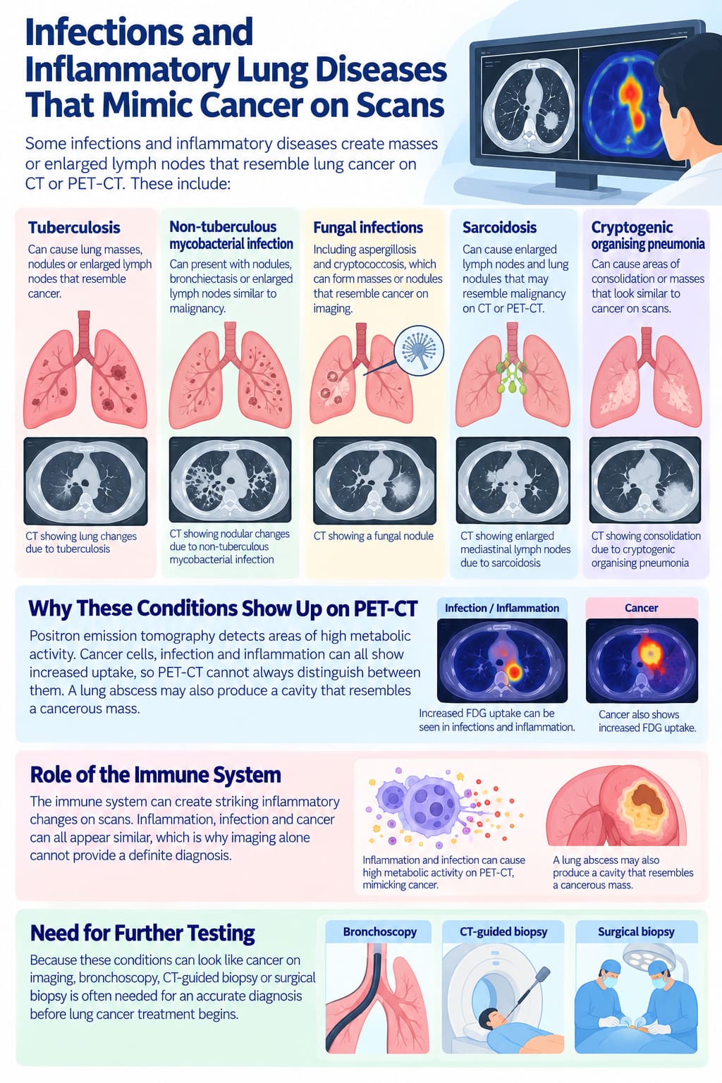
Some infections and inflammatory diseases create masses or enlarged lymph nodes that resemble lung cancer on CT or PET-CT. These include:
Tuberculosis
Non-tuberculous mycobacterial infection
Fungal infections, including aspergillosis and cryptococcosis
Sarcoidosis, which may resemble malignancy
Cryptogenic organising pneumonia
Positron emission tomography detects areas of high metabolic activity. Cancer cells, infection and inflammation can all show increased uptake, so PET-CT cannot always distinguish between them. A lung abscess may also produce a cavity that resembles a cancerous mass.
The immune system can create striking inflammatory changes on scans. For this reason, bronchoscopy, CT-guided biopsy or surgical biopsy is often needed for an accurate diagnosis before lung cancer treatment begins.
Heart Conditions, Pulmonary Embolism and Pulmonary Oedema
Heart conditions can produce breathlessness, cough and chest pain. Congestive heart failure commonly causes fatigue, fluid retention and trouble breathing. The National Heart, Lung, and Blood Institute identifies these symptoms as common reasons for hospital admission.
Pulmonary oedema is fluid in the lungs, usually caused by heart failure. It may cause sudden shortness of breath, pink frothy sputum and chest discomfort. A chest X-ray often shows diffuse shadowing rather than a single mass.
Pulmonary embolism is a blood clot in the lung. It may cause sudden breathlessness, sharp chest pain, coughing blood and a rapid heart rate. CT pulmonary angiography, ECG, blood tests and echocardiography can help separate these conditions from primary lung disease.
Mesothelioma and Other Chest Wall or Pleural Diseases
Mesothelioma is an asbestos-related cancer affecting the pleura, the lining around the lungs. Lung cancer begins in lung tissue. Both can cause mesothelioma symptoms such as chest pain, breathlessness, cough, fatigue, weight loss and pleural effusion.
Benign pleural plaques, pleurisy, empyema and asbestos-related pleural thickening can also mimic cancer. In a study of 311 cases initially diagnosed as mesothelioma, 111 were ultimately primary lung cancers.
CT and PET-CT can show pleural thickening or nodularity, but image-guided or thoracoscopic biopsy is usually needed to establish the diagnosis. Understanding the differences between lung cancer and mesothelioma is essential because treatment options differ.
Gastro-Oesophageal Reflux, Vocal Cord Problems and “Throat” Causes of Cough
A chronic cough may originate outside the lungs. Gastro-oesophageal reflux disease and laryngopharyngeal reflux can cause dry cough, hoarseness, throat clearing, abdominal pain and chest discomfort.
Vocal cord nodules, paralysis and throat cancer may also present with cough and hoarseness. If chest imaging is normal but symptoms persist, ENT assessment, reflux treatment or pH monitoring may be appropriate.
However, weight loss, coughing blood or a new abnormality on chest imaging should always be investigated.
How Doctors Differentiate Lung Cancer From Its Mimics
No single test confirms or excludes lung cancer. A lung cancer diagnosis combines symptoms, examination, risk factors, imaging tests and pathology.
Typical diagnostic tests include:
Chest X-ray for initial assessment
CT scans
PET-CT
Lung function tests
Blood tests and infection markers
Bronchoscopy, EBUS, CT-guided biopsy or surgical biopsy
Doctors consider age, cigarette smoking, asbestos exposure, speed of symptom onset, fever and infection markers. They also assess nodule growth and overall fitness before recommending surveillance, biopsy or surgery.
When an Initial Diagnosis Changes to Lung Cancer (or Away From It)
The initial diagnosis may be infection, COPD or musculoskeletal chest pain before lung cancer is recognised. This is particularly likely when early signs are subtle.
Typical scenarios include:
Recurrent pneumonia in the same lobe, including the left lung
A COPD flare-up that does not respond to usual treatment
Persistent chest pain after presumed strain
A suspicious lesion that proves benign after biopsy or surgery
Follow-up imaging is important after pneumonia, particularly for people over 50 with a smoking history. A normal chest X-ray does not exclude cancer, and CT is more sensitive when concern remains.
Multidisciplinary review and a second opinion from a thoracic specialist can support correct diagnosis, avoid overtreatment and improve disease control.
When to Seek Specialist Thoracic Surgical Advice
Seek specialist advice when imaging shows suspicious lung nodules, a mass or unexplained pleural fluid. Important warning signs include:
Persistent chest pain or breathing difficulties
Coughing blood
Unexplained weight loss
Lung shadows that do not resolve
Conflicting opinions about a lung lesion
Mr Marco Scarci can discuss further imaging, less invasive biopsy, keyhole lung surgery or surveillance. The aim is to confirm or exclude lung cancer safely, avoid unnecessary major surgery and establish an appropriate treatment plan.
Frequently Asked Questions
Can a normal chest X-ray completely rule out lung cancer?
No. Small tumours, lesions behind the heart or diaphragm and some upper-lobe cancers may not be visible on a standard chest X-ray. If symptoms persist or risk is higher, a CT scan is usually the appropriate next step.
How long should symptoms persist before I worry about lung cancer?
A cough lasting more than three weeks, new shortness of breath or chest pain that does not settle should be reviewed by a GP. Coughing blood, unexplained weight loss or repeated infections require prompt assessment, regardless of duration.
Do I still need tests if I have COPD or asthma and my symptoms just seem “a bit worse”?
Yes. A change from your normal symptoms may reflect infection, heart disease or developing lung cancer. New chest pain, coughing blood, night sweats or a persistent decline should not be assumed to be a routine flare-up, particularly in people over 50 or those with obstructive pulmonary disease COPD.
What is the difference between lung cancer and mesothelioma?
Lung cancer starts in lung tissue, while mesothelioma starts in the pleural lining around the lung and is strongly associated with asbestos exposure. Both may cause breathlessness, chest pain and fluid around the lungs. Imaging and biopsy are needed because treatment differs.
Can a benign lung nodule turn into cancer over time?
Most benign nodules, including granulomas and hamartomas, do not become cancer. However, new cancer can develop elsewhere in one or both lungs. Surveillance CT scans can confirm stability and guide a treatment plan.
